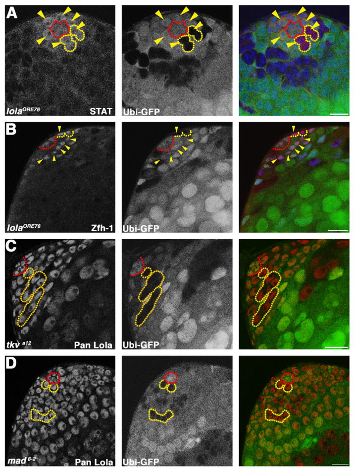
Fig. 3.

lola likely acts in parallel to the JAK-STAT and TGF-β pathways: (A) lolaORE76 mosaic testis stained with antibodies against STAT. Red: STAT. Green: Ubi-nls-GFP. Blue: DAPI. Red outline: hub. Yellow outlines: homozygous mutant GSC clones. Yellow arrowheads: GSCs positive for STAT. (B) lolaORE76 mosaic testis stained with antibodies against Zfh-1. Red: Zfh-1. Green: Ubi-nls-GFP. Blue: Tj. Red outline: hub. Yellow outlines: homozygous mutant CySC clones. Yellow arrowheads: CySCs expressing Zfh-1. (A) and (B) Scale bars: 15 μm. (C) and (D) tkva12 (C) and mad8–2, (D) mosaic testes at 3 dpci stained with pan Lola antibody. Red: Lola. Green: Ubi-nls-GFP. Red outline: hub. Yellow outlines: tkva12 (C) and mad8–2 (D) homozygous mutant GSCs and spermatogonial cysts. (C) and (D) Scale bars: 20 μm.
