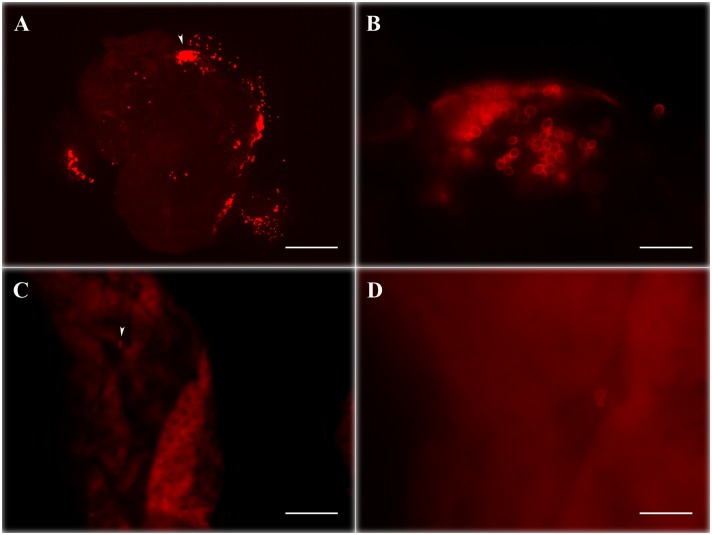
Figure 3. WaF17.12-KT fluorescence signal comparison in female mosquito midguts with or without yeast introduction.
Female mosquito midguts on the 10th day after feeding with sugar solution enriched with stimulated WaF17.12 (A and B) and sterile sugar solution (C and D). Red stained yeasts are abundantly visible in (A), while only few cells are detected in the control sample (C). Images B and D (bar = 20 µm) are magnification of samples shown in A and C respectively (bar = 200 µm) White arrow in A and C indicates the section of sample enlarged in B and D.

