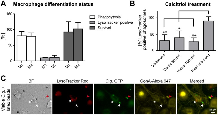
Figure 2. Phagosome maturation arrest occurs in different macrophage differentiation or activation states and is yeast phagosome-specific.

(A) Human M1-polarized and M2-polarized MDMs do not differ in central aspects of C. glabrata-macrophage interaction: phagocytosis, phagosome acidification and killing. Phagocytosis (MOI of 5) was quantified microscopically by determining the percentage of internalized (Concanavalin A stain-negative) yeasts out of total yeasts after 90 min. Phagosome acidification was quantified microscopically by determining the percentage of LysoTracker-positive phagosomes after 90 min. Survival of C. glabrata was determined by cfu-plating of macrophage lysates after 3 h of co-incubation and comparing to yeasts incubated without macrophages. (B) Treatment with vitamin D3 (calcitriol) has no influence on the number of LysoTracker-positive viable C. glabrata containing phagosomes of human MDMs. (C) MDMs co-infected with C. glabrata and latex beads show a acidification defect specific to C. glabrata containing phagosomes (LysoTracker-negative staining; white arrow) but acidify latex bead containing phagosomes (LysoTracker-positive staining; white asterisk). Representative image 90 min post infection. GFP-expressing C. glabrata is indicated in green and non-phagocytosed yeasts stained with Concanavalin A (ConA) are in yellow (marked with red arrows). Statistical analysis was performed comparing M1-type with M2-type macrophages (A) or drug treated with untreated viable C. glabrata (B) (n≥3; *p<0.05, **p<0.01 by unpaired Student’s t test).
