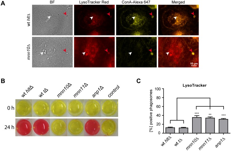
Figure 6. The influence of mannosyltransferases of C. glabrata on environmental alkalinization and acidification of phagosomes.
(A) Representative fluorescence microscopy images of wild type (wt hltΔ) C. glabrata and mnn10Δ mutant 90 min post infection, phagocytosed by human MDMs (left panels). LysoTracker staining is shown in red, while non-phagocytosed yeasts, stained with Concanavalin A (ConA), are shown in yellow. Phagocytosed yeasts are labeled with a white arrow while non-phagocytosed yeasts are marked with red arrows. (B) Co-localization with LysoTracker was quantified for phagosomes containing wild type (wt hltΔ or wt tΔ) or mutant (mnn10Δ, mnn11Δ, anp1Δ) C. glabrata at 90 min post infection. Statistical analysis was performed comparing mutant with wild type C. glabrata (n≥3; **p<0.01, ***p<0.005 by unpaired Student’s t test). (C) mnn10Δ and mnn11Δ mutants showed severe defects in environmental alkalinization in vitro, while alkalinization by the anp1Δ mutant was comparable to isogenic wild type levels. 1×10⁶ C. glabrata cells/ml were inoculated in a 24 well plate with liquid YNB medium with 1% casamino acids and 20 mg/l phenol red and incubated for 24 h.

