
Figure 3. Schematic presentation of the HOXD13 structure and sequences alignment analysis.
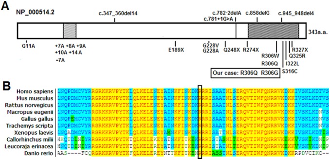
A: Schematic presentation of the HOXD13 structure and annotated mutations identified in families. B: Partial amino acid sequences alignment in homeodomain of HOXD13 among several species. The position of mutated amino acid of our cases is indicated by the black box.