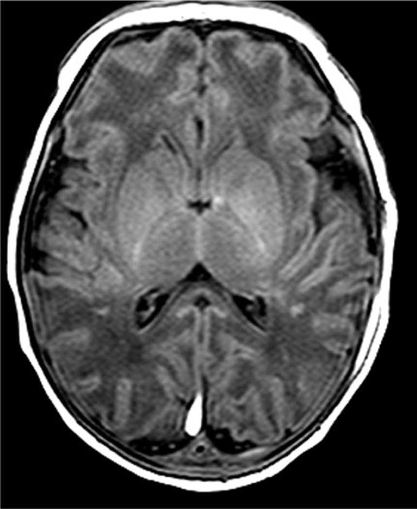
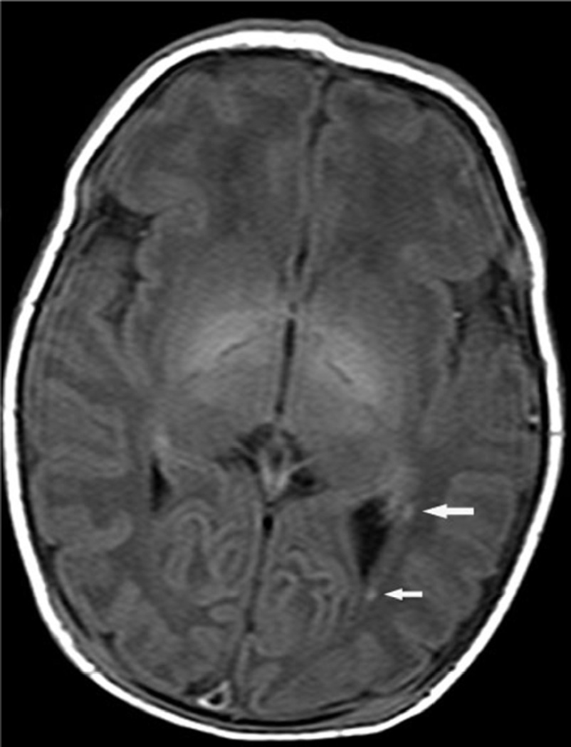
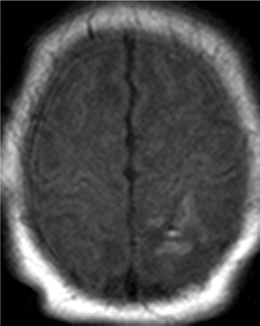
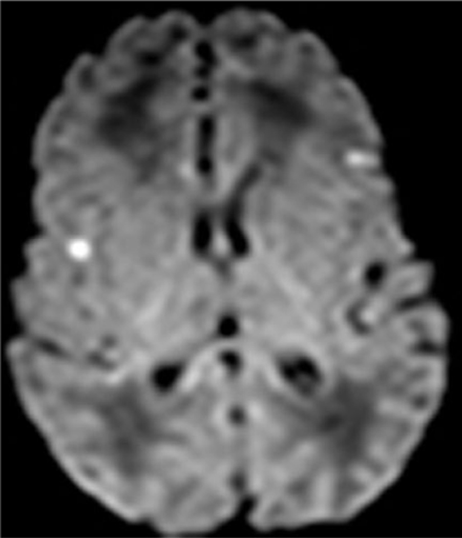
Fig. 1.
Examples of MRI showing minimal brain injury (score=1). White matter lesions most often involved the deep parietal white matter and tended to be bilateral.
a. Axial T1 image of a 7 day old infant shows multiple punctate areas of T1 shortening within the parietal white matter. The basal ganglia and thalami are normal
b. White matter lesions (arrows) were occasionally unilateral.
c. Isolated focal cortical infarct in 39 week infant imaged at day of life 7.
d. In another patient, there are scattered areas of restricted diffusion within the cerebral cortex.


