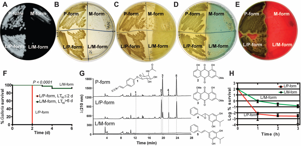
Fig. 3.
Change in phenotypes, pathogenicity, secondary metabolite production, and persistence between P and M forms. (A) to (E) Quadrants on Petri dishes containing the P form (left), M form (right), unlocked forms (top), and locked forms (bottom). (A) Bioluminescence produced by the P form, which is absent in the M form (note that bioluminescence from P-form revertants is visible in the M form). (B) The P form is yellow and opaque, and the M form is unpigmented and transparent. (C) Antimicrobial activity produced at 48 hours of growth on LBP by the P form and not the M form against a softagar overlay containing Micrococcus luteus indicator bacteria. (D) Production of siderophore iron-chelating activity by the P form and not the M form indicated as a zone of clearing as iron is removed from chrome azural S chelator. (E) Hemolytic activity on sheep blood agar produced by the P form but not the M form. (F) Virulence of the L/P form and not the L/M form after injection into Galleria mellonella. (G) Metabolite analysis detected the production of rhabduscin (1), two anthraquinone pigment molecules (2 and 3) and two hydroxystilbene molecules (4 and 5), which were mostly absent in the L/M form. (H) There are 50 times more persister cells tolerant to ciprofloxacin in the L/M form than in the L/P form.

