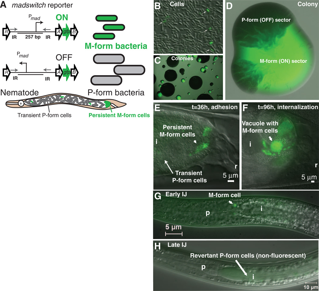
Fig. 4.
Single-cell reporter studies of the madswitch during the development of symbiosis in nematodes. (A) A gfp gene was inserted between madA and madB so as not to disrupt expression of downstream mad genes. The madswitch ON cells are small, green fluorescent cells capable of maternal adhesion, and the madswitch OFF cells are large, non-fluorescent cells that are the majority of cells transiently present in the maternal nematode intestine. (B) Few cells (white arrows) have the madswitch oriented ON in culture. (C) Small colonies have the madswitch oriented ON, and large colonies have the madswitch oriented OFF. (D) An isolated colony of the M form (green) develops dark, opaque sectors that are madswitch oriented OFF P form. (E) madswitch-ON cells adhere to the posterior maternal nematode intestine, and most cells transiently present are not fluorescent, with the madswitch OFF. (F) Most adherent cells that invade and grow inside vacuoles of the rectal gland cells have the madswitch oriented ON. (G) One or two cells on or inside the pharyngeal intestinal valve cells have the madswitch oriented ON. (H) Seven days after the symbionts fully colonize the IJs, all cells are not fluorescent with the madswitch OFF, and again, the insect pathogenic P form is arming the nematode for insect infection. i, intestine; r rectum; p, pharynx.

