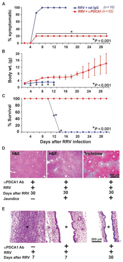
Fig. 6.
Prevention of biliary atresia by depletion of pDCs. (A to C) Improvements in symptoms (A), weight gain (B), and survival (C) for RRV-infected neonatal mice receiving anti-PDCA1 antibody (Ab) (red) (n = 22) or IgG isotype control antibody (blue) (n = 10). (D) Liver sections stained with hematoxylin and eosin (H&E) show that mice with symptoms had portal expansion and inflammation (middle panel) with collagen deposition (right panel), whereas asymptomatic mice had normal liver histology (left panel). (E) H&E staining of longitudinal sections of extrahepatic bile duct shows lumenal obstruction by inflammatory cells in mice receiving IgG isotype antibody (asterisks). In contrast, mice receiving anti-PDCA1 antibody demonstrate a lack of lumenal inflammation and intact bile duct epithelium.

