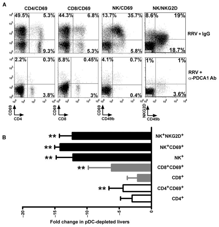
Fig. 7.
Decreased hepatic lymphocytes in RRV-infected mice in response to pDC depletion. Flow cytometric analyses of activation markers of CD4+, CD8+, and NK cells 7 days after the injection of RRV (with and without injections of anti-PDCA1 antibodies) into newborn mice. (A) Representative dot plots; values in each quadrant represent percent cells positive for respective cell surface markers. (B) Fold change for expression of activation markers by individual populations of hepatic lymphocytes from mice infected with RRV and injected with either anti-PDCA1 antibody or IgG isotype antibody control. Data are representative results of three experiments. **P < 0.01 (Mann-Whitney test) when compared to isotype control.

