. 2014 Mar;30(1):1–5. doi: 10.5487/TR.2014.30.1.001

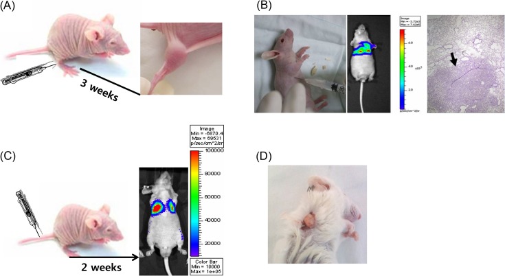
Fig. 1. Various xenograft models. (A) Ectopic xenograft model. The cancer cells were subcutaneously injected into Balb/c nude mice. After approximately two weeks, the tumor was observed. (B) Orthotopic xenograft model. Human non-small cell lung cancer cells (A549 cells) were injected into the thoracic cavity of Balb/c nude mice. Tumor was observed by in vivo optical imaging. Isolated lung tissue was stained and observed by microscopy. (C) Metastasis model. Luciferase-expressing cancer cells were injected into the tail vein. Tumor was observed by in vivo optical imaging. (D) Patient-derived tumor xenograft model. Patient-derived tumor tissues were transplanted into the SCID mouse.

Fig. 1.