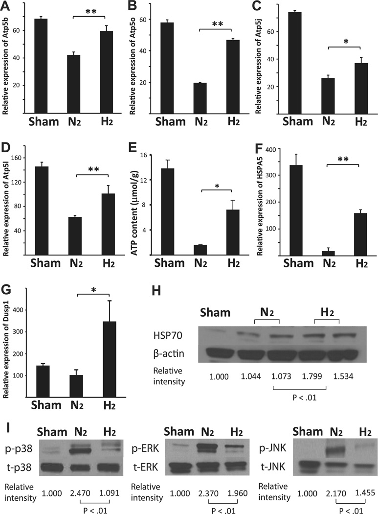
Fig. 3.
Quantitative RT-PCR for (A) ATP5b, (B) ATP5o, (C) ATP5j and (D) ATP5 l (n = 4 for each group; **p < 0.01 and *p < 0.05). (E) Lung ATP content prior to implantation. (n = 4 per group *p < 0.05). Quantitative RT-PCR for (F) HSPA5 and (G) DUSP1 (n = 4 for each group; **p < 0.01 and *p < 0.05). (H) Western blots for HSP70 and β-actin on protein extracts from lung grafts taken after 3 h of mechanical ventilation and 4 h of cold storage. (I) Western blots for phosphorylated (p) and total (t) p38, ERK1/2 and JNK on protein extracts from lung grafts taken 2 h after reperfusion. The images are representative of 3 independent experiments (n = 3 for each group).

