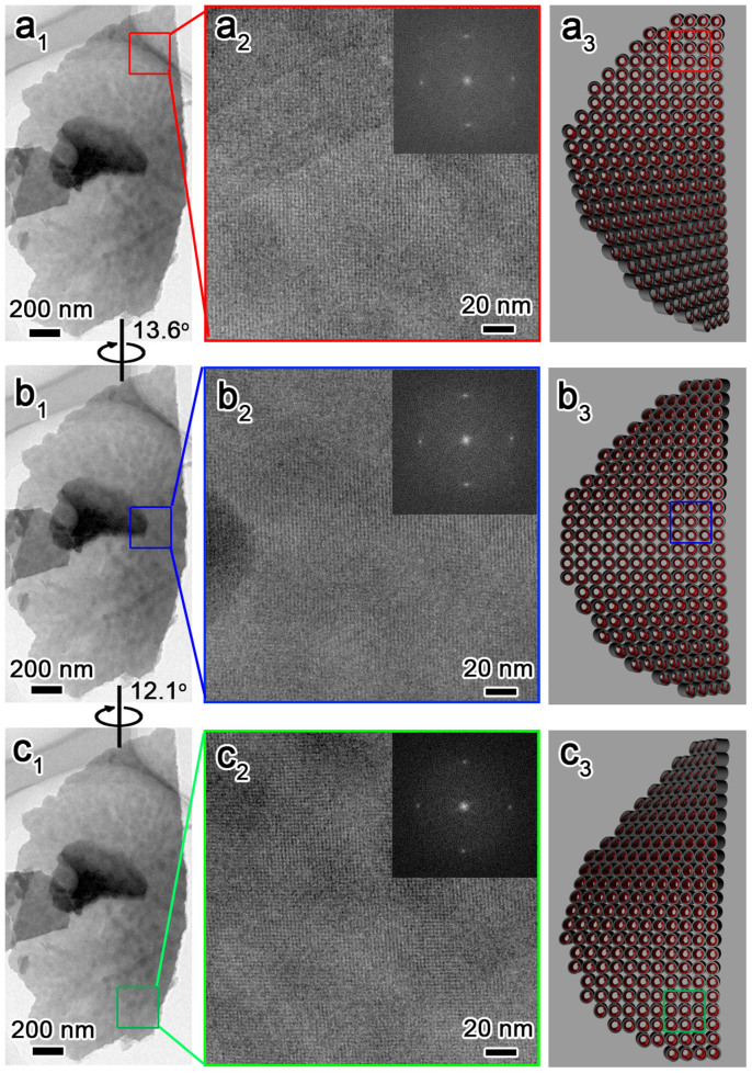
Figure 3. Microscopic helical structures of DNA chiral packing, macroscopic helical morphologies of the twisted blades and the corresponding schematic representation of the CDSFs shown in Figure 1a.
The blades were bent, and the 2D-square p4mm lattice aligned with the incident electron beam in the top region (a1–a3). We aligned the middle (b1–b3) and bottom (c1–c3) portions by tilting the sample in a clockwise manner along its (10) axes by 13.6° and then 12.1°, which unambiguously indicated left-handed DNA chiral packing and left-handed helical blades.

