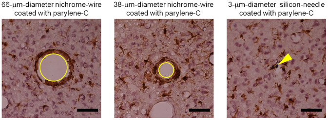
Figure 6. Immunohistochemical evaluation of tissue four days after implantation of nichrome wires and a silicon microneedle.

Microglia (brown) and nucleus (violet) are identified with immunohistochemical staining with the Iba1 antibody and the counterstaining with hematoxylin, respectively. Panels show microglia around a 66-μm-diameter nichrome wire (left), a 38-μm-diameter nichrome wire (center), and a 3-μm-diameter silicon needle (right). All of the wires and the needle were coated with parylene-C (thickness of 1 μm). Yellow circles in the left and center panels show traces of the 66-μm-diameter and 38-μm-diameter nichrome wires, respectively. Yellow arrow in the right panel shows the silicon microneedle remaining in the tissue. While the length of the silicon microneedle was 750 μm and those of the nichrome wires were 1 mm, these tangentially sliced sections were sampled at depths between 320 μm and 440 μm from the cortical surface of a rat. Scale bar: 50 μm.
