
Figure 4. Primary blast induced oxidative and nitrosative stress in the rat brain microvessels.
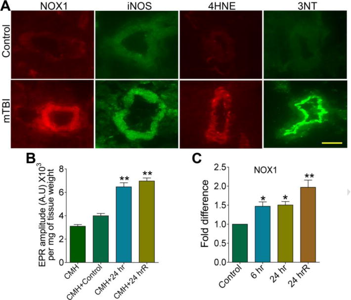
(A) Immunofluorescent staining of NOX1, iNOS, 4HNE, and 3NT in intact brain microvessels of rats subjected to a single blast with 123 kPa peak overpressure, 24 hours post-exposure. (B) ROS generation was detected by electron paramagnetic resonance (EPR) in brain tissue slices from 24 hr and 24 hrR post-exposure of mTBI of 123 kPa blast exposed and compared with control. Results are expressed in EPR amplitude arbitrary units per milligrams of tissue weight. (C) Changes in mRNA level of NOX1 in brain cortical tissues of rats in different time intervals of 123 kPa blast post-exposure by quantitative RT-PCR using TaqMan primers. Values are mean ±SEM (n = 3 in B and C). Statistically significant *p<0.05; **p<0.01 versus control in C; and versus CMH+control in B. Scale bar: 5 μm in all panels of A.