Abstract
Aim:
Peroxisome proliferator-activated receptor gamma (PPARγ) is a therapeutic target for obesity, cancer and diabetes mellitus. In order to develop potent lead compounds for obesity treatment, we screened a natural product library for novel PPARγ antagonists with inhibitory effects on adipocyte differentiation.
Methods:
Surface plasmon resonance (SPR) technology and cell-based transactivation assay were used to screen for PPARγ antagonists. To investigate the antagonistic mechanism of the active compound, we measured its effect on PPARγ/RXRα heterodimerization and PPARγ co-activator recruitment using yeast two-hybrid assay, Gal4/UAS cell-based assay and SPR based assay. The 3T3-L1 cell differentiation assay was used to evaluate the effect of the active compound on adipocyte differentiation.
Results:
A new thiophene-acetylene type of natural product, 7-chloroarctinone-b (CAB), isolated from the roots of Rhaponticum uniflorum, was discovered as a novel PPARγ antagonist capable of inhibiting rosiglitazone-induced PPARγ transcriptional activity. SPR analysis suggested that CAB bound tightly to PPARγ and considerably antagonized the potent PPARγ agonist rosiglitazone-stimulated PPARγ-LBD/RXRα-LBD binding. Gal4/UAS and yeast two-hybrid assays were used to evaluate the antagonistic activity of CAB on rosiglitazone-induced recruitment of the coactivator for PPARγ. CAB could efficiently antagonize both hormone and rosiglitazone-induced adipocyte differentiation in cell culture.
Conclusion:
CAB shows antagonistic activity to PPARγ and can block the adipocyte differentiation, indicating it may be of potential use as a lead therapeutic compound for obesity.
Keywords: peroxisome proliferator-activated receptor, antagonist, surface plasmon resonance, recruitment of the coactivator, adipocyte differentiation
Introduction
The peroxisome proliferator-activated receptors (PPARs) belong to a superfamily of nuclear hormone receptor and function as heterodimers with the retinoid X receptor (RXR) to regulate the transcription of their target genes via binding to specific peroxisome proliferator response elements (PPRE)1. There are three different PPAR genes that encode four distinct proteins: PPARα, PPARδ, PPARγ1, and PPARγ22, 3. Among them, PPARγ is expressed at highest levels in adipose tissue and lower levels in several other tissues. It plays a key role in many metabolic processes, including insulin sensitivity and adipocyte differentiation4, 5. PPARγ can be activated by arachidonic acid-metabolites6, 7, 8 and fatty acid-derived components9.
Currently, obesity is considered as the most common metabolic disease in developed nations. In 2003–2006, the US body mass index growth charts showed that 11.3% of US children and adolescents aged 2 through 19 years old were at or above the 97th percentile, and 31.9% were at or above the 85th percentile10. These types of statistics provide urgent impetus for the development of efficient strategies focused onto reducing the obesity epidemic. PPARγ is one of the important therapeutic targets against obesity. Adipocyte differentiation appears to be controlled by two major factors or groups of factors: PPARγ and the C/EBPs. PPARγ is a component of the adipogenic transcription factor ARF611. C/EBPα promotes the adipogenic program, while expression and activation of PPARγ efficiently induce adipogenesis12, thereby confirming that PPARγ and the C/EBPs play important roles in terminal adipocyte differentiation13. Thiazolidinediones (TZDs), a class of insulin-sensitizing agents, bind to and activate PPARγ. However, an unwanted side effect of TZD treatment is modest weight gain partly caused by increased adipogenesis and the influx of fatty acids into adipose tissue. It has been revealed that the antagonist or partial agonist of PPARγ might demonstrate promising applications in the discovery of novel anti-diabetic agents that may retain efficacious insulin sensitizing properties and minimize the potential side effects. For example, the published antagonists of PPARγ such as BADGE14, PD06823515, T007090716, and SR-20217, exhibited the effect of blocking adipocyte differentiation. Notably, SR-20217 could improve the insulin sensitivity and reduce glucose levels of plasma, implying its potential application in the treatment of obesity and type 2 diabetes.
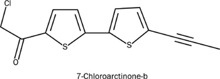
The natural product 7-chloroarctinone-b (CAB, Figure 1) is a new thiophene-acetylene type of derivative isolated from the Rhaponticum uniflorum's roots18. Rhaponticum uniflorum (Asteraceae family) is a perennial herbaceous plant which is widely distributed in the northern part of China. Thiophene-acetylenes (ie ethynylthiophenes) represent a unique class of natural products exhibiting a wide variety of biological activities ranging from antitumor, antiviral, anti-HIV, antifungal to insecticidal activities 18.
Figure 1.
Structure of CAB (7-chloroarctinone-b).
By random screening against our lab in-house natural product library, CAB was discovered as a new PPARγ antagonist. The CAB antagonistic activity against the rosiglitazone-induced recruitment of the coactivator for PPARγ was evaluated in both Gal4/UAS and yeast two-hybrid systems. CAB could efficiently antagonize both hormone and rosiglitazone induced adipocyte differentiation in cell culture. It is thus expected that CAB might be potentially used as a lead compound for anti-obesity agent discovery.
Materials and methods
Reagents
Rosiglitazone and AP2 antibody were obtained from Cayman Chem Co (Ann Arbor, MI, USA). GW9662 was obtained from Merck. Yeast nitrogen base without amino acids, yeast synthetic drop-out medium supplement without tryptophan, yeast synthetic drop-out medium supplement without leucine and tryptophan, D-(+)-glucose, and p-nitrophenyl a-D-galactopyranoside (PNP-α-Gal) were obtained from Sigma Chemical Co. All cell culture reagents were obtained from GIBCO. Lipofectamine-2000 was obtained from Invitrogen. GAPDH antibody was obtained from Kangcheng Bio-tech (Shanghai, China). All other solvents and reagents were purchased commercially and used without further purifications.
Plasmids
The yeast expression vectors pGBKT7 and pGADT7 were generously provided by Prof Y GONG (Shanghai Institutes for Biological Sciences, CAS, China). CMV-mCBP (mouse CBP) was from Prof MG ROSENFELD (Howard Hughes Medical Institute, University of California, USA). pET15bhPPARγ-LBD was kindly donated by Dr J UPPENBERG (Department of Structural Chemistry, Pharmacia and Upjohn, Sweden). pcDNA3.1-hPPARγ was a gift from Dr X GAO (Chengdu Institute of Biology, CAS, China). The reporter gene pSV-PPRE-Luc was kindly provided by Dr Ronald M Evans (The Salk Institute for Biological Studies, La Jolla, CA, USA). Gal4-PPARγ-LBD expression plasmid and UASE1b-TATA-Luc reporter gene were generously donated by Prof J JAMESON (Department of Medicine, Northwestern Memorial Hospital). PPARα-LBD and PPARδ-LBD were amplified by PCR from pSG-hPPARα (provided by Dr X LU, Shenzhen Chipscreen Biosciences Ltd) and pAdTrack-PPARδ (provided by Dr B VOGELSTEIN, Howard Hughes Medical Institute, US), respectively, and then subcloned into vector pET15b to express the His-tagged fusion proteins. The DNA coding for RXRα-LBD (aa 223–462) was subcloned into vector pET22b.
Protein preparation
PPARγ-LBD, PPARα-LBD, PPARδ-LBD, and RXRα-LBD (aa 223–462) were prepared according to our reported methods19. Protein concentration was measured by the standard Bradford method.
SPR measurements
Binding affinity of CAB towards PPARγ-LBD, PPARα-LBD, PPARδ-LBD, or RXRα-LBD was assayed by SPR technology based Biacore 3000 instrument (Biacore AB, Uppsala, Sweden). The proteins to be covalently bound to the chips were diluted into 10 mmol/L sodium acetate buffer (pH 4.5) to a final concentration of 0.10 mg/mL and then immobilized to CM5 chips using a standard amine-coupling procedure. Prior to the start of experimentation, baseline was equilibrated with a continuous flow of running buffer [10 mmol/L HEPES, 150 mmol/L NaCl, 3 mmol/L EDTA, and 0.005% (v/v) surfactant P20, pH 7.4]. Different concentrations of CAB were then injected into the channels at a flow rate of 20 μL/min for 120 s, followed by disassociation for 150 s. The 1:1 Langmuir binding fitting model was used to determine the equilibrium dissociation constant (KD) of compounds.
In the investigation of the PPARγ-LBD/RXRα-LBD interaction, purified RXRα-LBD protein was immobilized on a CM5 sensor chip, and the published approach was then applied to test the binding ability of PPARγ-LBD towards RXRα-LBD in the presence or absence of compounds19. All the results were reflected by RU (resonance units) values recorded directly by the Biacore 3000 instrument.
Cell culture
HEK293T (human embryonic kidney) cells were cultured in Delbecco's modified Eagle's medium (DMEM) supplemented with 10% FBS. 3T3-L1 cells were cultured in DMEM supplemented with 10% FBS, biotin (8 mg/L) and Ca-pantothenate (4 mg/L). All cells were cultured at 37 °C in a humidified atmosphere with 5% CO2.
Yeast two-hybrid assay
Yeast cells (strain AH109) were transformed with plasmids pGADT7-mCBP (aa 1–464) and pGBKT7-PPARγ-LBD (aa 204-447). The transformed yeast cells were diluted to an initial OD600 value of 0.3−0.4 and incubated with vehicle (DMSO) or the tested compound. After 24-h incubation, the α-galactosidase activities were evaluated following the previous published approach protocol19.
Transfection and luciferase assays
Transient transfection was carried out in 24-well plates. Cells were transfected in opti-MEM with the transfection reagent Lipofectamine-2000 (Invitrogen, USA) according to the manufacturer's protocols. The renilla vector pRL-SV40 (50 ng/well) was used to normalize the transfection efficiency. After a 5-h transfection, the medium was replaced with fresh DMEM supplemented with 10% FBS, and the cells were incubated with vehicle (DMSO) or compound(s) for another 18 h. Finally, cells were lysed and luciferase activities were measured using Dual Luciferase Assay System kit.
The mammalian one-hybrid assay system was used to investigate the effects of CAB on PPARγ-LBD in HEK-293T cells, where the UAS-TK-Luc reporter and the fusion construct of GAL4-PPARγ-LBD were transiently co-transfected. In addition, the effects of CAB on the full-length PPARγ were also examined in HEK-293T cells that were transiently transfected with plamids of pSV-PPRE-Luc (0.3 μg/well), pCDNA3.1-PPARγ (0.2 μg/well) and pCDNA3.1-RXRα (0.2 μg/well)20.
3T3-L1 cell differentiation assays
To determine whether CAB can antagonize rosiglitazone-induced adipocyte differentiation, 3T3-L1 preadipocytes were cultured as described and induced to differentiate by treatment with rosiglitazone (1 μmol/L) in the presence or absence of CAB for 6 days. To determine whether CAB is antagonistic on hormone-induced adipocyte differentiation, 3T3-L1 cells were stimulated with an MDI cocktail [0.115 mg/L methylisobutylxanthine (MIX), 0.39 mg/L dexamethasone (DEX) and 1 mg/L insulin] and CAB for 3 days, and medium was then changed to fresh medium containing 1 mg/L insulin and CAB for another 3 days.
Adipogenesis was determined by staining lipids with Oil Red O. The expression of adipocyte marker (aP2) was determined by Western blot analysis using anti-aP2 antibody (Cayman chemical; diluted 1:200).
Results
CAB is a PPARγ antagonist
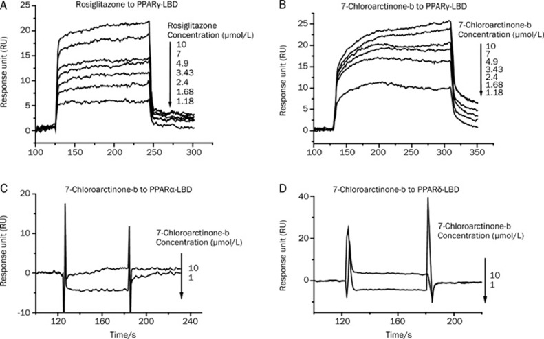
CAB was initially identified in a screen for compounds that bound to PPARγ-LBD. SPR technology based Biacore 3000 instrument was used to carry out the kinetic analysis and selectivity of CAB binding to PPARγ-LBD. The equilibrium dissociation constant (KD) of PPARγ binding to CAB or rosiglitazone (as a positive control) was obtained by fitting the sensorgrams with the 1:1 (Langmuir) binding fit model. As shown in Figure 2B, CAB showed high binding affinity to PPARγ-LBD (KD=2.63 μmol/L, Table 1), similar to that of rosiglitazone (KD=2.83 μmol/L, Figure 2A, Table 1). Moreover, to further inspect the potential binding specificity of CAB to PPARγ, CAB binding to PPARα-LBD and PPARδ-LBD was also examined. The SPR results clearly indicated that CAB has no affinity to PPARα-LBD or PPARδ-LBD (Figure 2C and 2D), suggesting that CAB is a selective ligand of PPARγ.
Figure 2.
CAB exhibited a highly specific binding affinity against PPARγ as evaluated by SPR analysis. The sensorgrams were obtained from injection of series of concentrations of (A) rosiglitazone or (B) CAB over the immobilized PPARγ-LBD surface or from injection of series of concentrations of CAB over immobilized (C) PPARα-LBD, (D) PPARδ-LBD surface. The ligands were injected for 120 s, and dissociation was monitored for more than 150 s. The concentrations (μmol/L) are shown next to the arrows.
Table 1. SPR technology based kinetic analysis for 7-chloroarctione-b (CAB) or rosiglitazone/PPARγ-LBD interaction.
| kon (mol·L−1·s−1) | koff (s−1) | KD (μmol/L) | χ2 | |
|---|---|---|---|---|
| Rosiglitazone | 4.35×10⁴ | 0.123 | 2.83 | 1.25 |
| CAB | 1.23×10³ | 3.24×10⁻³ | 2.63 | 0.703 |
kon, association rate constant; koff, dissociation rate constant; KD, equilibrium dissociation constant; χ2, statistical value in Biacore.
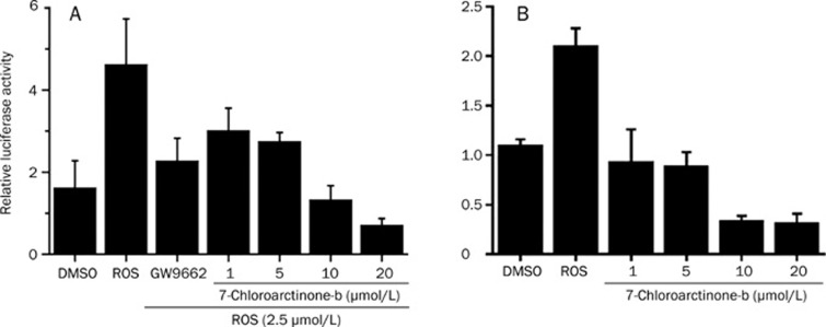
To further investigate the ability of CAB in modulation of PPARγ transcription, transactivation assay was performed. In the assay, HEK-293T cells were cotransfected with full-length PPARγ, RXRα, and a reporter plasmid containing the PPAR response element. Transfected cells were incubated with different concentrations of CAB with or without rosiglitazone. As shown in Figure 3A, co-treatment with 2.5 μmol/L rosiglitazone and increasing concentrations of CAB resulted in a dose-dependent decrease in rosiglitazone-stimulated PPARγ transcriptional activity. As shown in Figure 3B, CAB itself could also inhibit PPARγ basal transcriptional activity induced by endogenous PPARγ ligands. These results indicate that CAB is a dose-dependent inhibitor of PPARγ transcriptional activity whether stimulated by its agonist, rosiglitazone, or by other ligands.
Figure 3.
CAB inhibited PPARγ transcriptional activity as evaluated by transactivation assay. HEK293T cells were co-transfected with PPARγ, RXRα and reporter gene, while the control plasmids were treated (A) with or (B) without rosiglitazone (2.5 μmol/L) and increasing concentrations of CAB for 18 h. The cells were harvested for luciferase and β-galactosidase assays. The luciferase activity was normalized with β-galactosidase activity. The concentration of GW9662 (PPARγ antagonist) was 1 μmol/L.
SPR based investigation of CAB in antagonizing PPARγ/RXRα heterodimerization
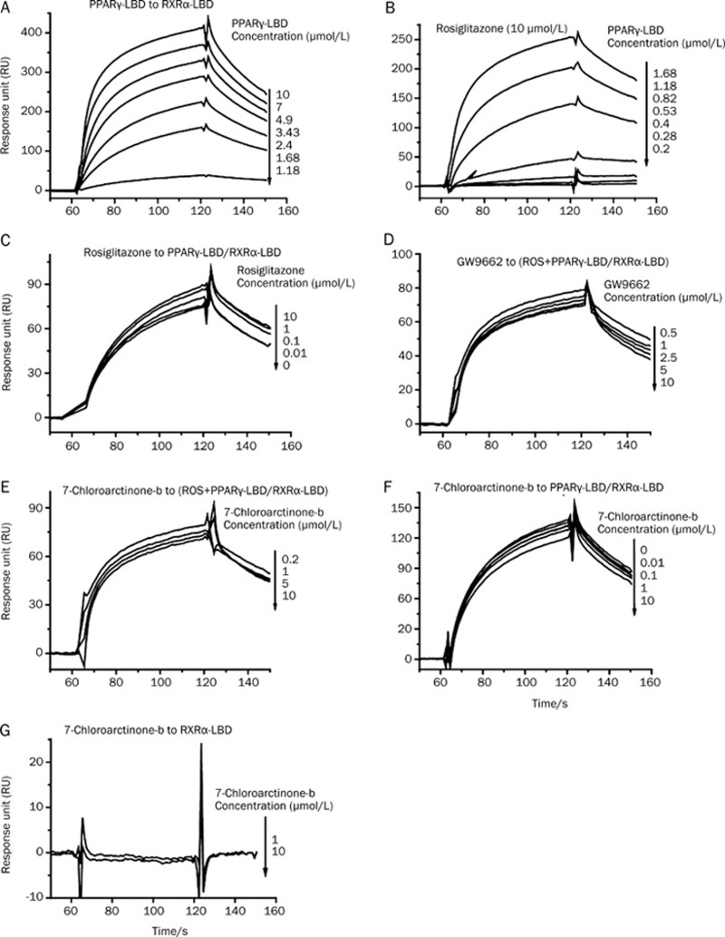
As reported, PPARγ formed heterodimer with the nuclear receptor RXR before binding to the specific PPRE to regulate transcription of their target genes2. We thus used SPR technology to test the potential effects of CAB on PPARγ-RXRα dimerization. PPARγ-LBD/RXRα-LBD binding was confirmed as shown in Figure 4A, while the effects of PPARγ agonist rosiglitazone and antagonist GW9662 on PPARγ/RXRα dimerization as positive controls were inspected (Figure 4B–4D). Rosiglitazone is the positive control in the experiment of testing the effect on inducing the PPARγ-RXRα dimerization, while GW9662 is the positive control in the experiment of testing the effect on restraining the PPARγ-RXRα dimerization stimulated by rosiglitazone. As expected, rosiglitazone could stimulate PPARγ/RXRα interaction, whereas GW9662 restrained such stimulation. As shown in Figures 4E and 4F, in either group with or without rosiglitazone, CAB substantially decreased the affinity of PPARγ to RXRα, but had no binding to RXRα (Figure 4G). Therefore, combining all the above-mentioned results, we thereby concluded that CAB was able to antagonize PPARγ-LBD/RXRα-LBD dimerization by specifically binding to PPARγ-LBD.
Figure 4.
CAB antagonized PPARγ/RXRα heterodimerization as evaluated by SPR analysis. The sensorgrams were obtained by injection of series of concentrations of PPARγ-LBD over the immobilized RXRα-LBD surface with (A) DMSO, (B) rosiglitazone (10 μmol/L), or by injection of PPARγ-LBD (0.3 μmol/L) incubated with series of concentrations of (C) rosiglitazone, (D) GW9662, or series of concentrations of CAB with (E) or without (F) rosiglitazone (10 μmol/L), or by injection of series of concentrations of CAB over the immobilized RXRα-LBD surface (G).
CAB inhibited the recruitment of PPARγ coactivator
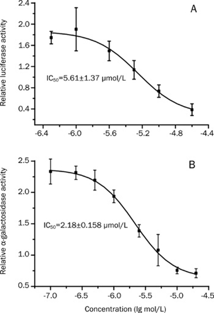
To investigate the effect of CAB on the ability to recruit the transcription coactivator of PPARγ at cell level, we co-transfected the UAS-TK-Luc reporter and the fusion construct of GAL4-PPARγ-LBD into HEK-293T cells. As shown in Figure 5A, CAB could efficiently inhibit rosiglitazone's (5 μmol/L) agonistic effects with an IC50 of 5.61 μmol/L. We postulated that CAB might have blocked the binding of PPARγ-LBD to its coactivator.
Figure 5.
CAB inhibited the recruitment of PPARγ coactivator. (A) Dose-dependent effects of CAB on the ability of PPARγ coactivator recruitment as evaluated by UAS/Gal4 system. HEK293T cells were co-transfected with Gal4-PPARγ-LBD and the UASE1b-TATA-Luc reporter gene, while the control plasmids were treated with rosiglitazone (0.5 μmol/L) and increasing concentrations of CAB for 18 h. The cells were harvested for luciferase and β-galactosidase assays. The values shown are the means±standard deviations from three independent experiments. (B) Dose-dependent effects of CAB on the recruitment of PPARγ transcription co-activator CBP as determined by yeast two-hybrid system. The overnight cultures of yeast cells containing pGADT7-CBP and pGBKT7-PPARγ were diluted with fresh media to an initial OD600 of 0.3 and treated with rosiglitazone (10 μmol/L) and increasing concentrations of CAB or DMSO (as vehicle control) for 16 h at 30 °C. Relative α-galactosidase activity was determined by OD410/OD600. The values shown are the means±standard deviations from three independent experiments.
To further confirm the above hypothesis, the yeast two-hybrid system regarding the PPARγ-LBD and its co-activator CBP (cAMP-response-element binding protein) was constructed. CBP is a critical transcription co-activator of PPARγ, and its interaction with PPARγ could be enhanced by the binding of rosiglitazone to PPARγ21. To test the antagonistic activity of CAB on this rosiglitazone-enhanced PPARγ/CBP binding, we transformed pGADT7-mCBP (aa 1–464) and pGBKT7-PPARγ-LBD (aa 204–447) plasmids into the yeast strain AH109 and investigated their interactions with or without compounds. As shown in Figure 5B, CAB could efficiently antagonize the rosiglitazone (10 μmol/L) stimulated PPARγ-LBD/CBP interaction with an IC50 of 2.18 μmol/L, consistent with the result (IC50=5.61 μmol/L) from the UAS/Gal4 system based assay.
CAB could efficiently block the differentiation of adipocyte 3T3-L1
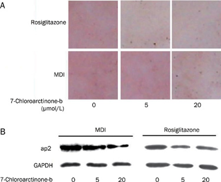
PPARγ is predominantly expressed in adipose tissue, where it plays critical roles in adipocyte differentiation and fat deposition. Since PPARγ antagonists have ever been reported to be able to inhibit adipocyte differentiation, we then investigated whether CAB could also prevent adipogenesis induced by the standard adipogenic mixture. Confluent 3T3-L1 preadipocytes were co-treated with a standard adipogenic mixture containing dexamethasone, IBMX, and insulin in the presence of increasing concentrations of CAB. After a 6-day incubation, adipocyte differentiation was visually evaluated by Oil Red O staining for lipid content and by measuring the expression of the adipocyte differentiation marker aP2. CAB could significantly inhibit hormone-induced 3T3-L1 adipocyte differentiation in a dose-dependent manner, as reflected by the decrease in lipid content revealed by Oil Red O staining and by the reduced expression of the adipocyte differentiation marker, aP2 (Figure 6A and 6B).
Figure 6.
CAB inhibited 3T3-L1 adipocyte differentiation. (A) Confluent 3T3-L1 preadipocytes were co-treated with 1 μmol/L rosiglitazone in the presence of increasing concentrations of CAB for 6 days or MDI stimulus (0.115 mg/L methylisobutylxanthine (MIX), 0.39 mg/L dexamthasone (DEX) and 1 mg/L insulin) and CAB for 3 days, and medium was then changed to fresh medium containing 1 mg/L insulin and CAB for another 3-day. Adipogenesis was determined by staining of lipids with Oil Red. (B) Western blot analysis of aP2 in 3T3-L1 preadipocytes treated with the indicated compounds.
To further confirm CAB as a PPARγ antagonist to block 3T3-L1 cell differentiation, the ability of CAB in inhibiting rosiglitazone-activated adipogenesis was also examined. As indicated in Figure 6A and 6B, similar to the case in the inhibition of hormone-induced 3T3-L1 adipocyte differentiation, CAB could also restrain the rosiglitazone-induced adipogenesis.
Discussion
In the current work, the natural product 7-chloroarctinone-b (CAB) was randomly screened out as a novel PPARγ antagonist from our lab in-house natural product library. CAB was isolated from the roots of Rhaponticum uniflorum (L.) DC., and exhibited a wide variety of biological activities including antitumor, antiviral, anti-HIV, antifungal and insecticidal activity18. SPR technology based investigation and transactivation assay demonstrated that CAB was a specific PPARγ antagonist. To further examine the potential antagonistic mechanism of this compound, its effects on PPARγ/RXRα heterodimerization and PPARγ co-activator recruitment were inspected. The results indicated that CAB considerably antagonized both rosiglitazone-induced PPARγ-LBD/RXRα-LBD binding and rosiglitazone-simulated PPARγ coactivator recruitment.
As previously reported, there are at least two pathways involved in 3T3-L1 adipocyte differentiation. One involves PPARγ and the other C/EBP22. PPARγ and C/EBPs are both known to be the direct transcriptional activators of several fat cell genes, and the best characterized adipocyte-specific regulatory DNA sites contain the binding sites for both factors23. Apart from C/EBPα, ectopic expression of C/EBP β and -δ can also induce the adipocyte differentiation of fibroblasts24. It has been proposed that PPARγ and C/EBPα could synergize each other to powerfully promote the adipocyte developmental program in fibroblastic cells. The PPARγ pathway exists in various tissues in addition to adipose and is targeted for therapeutic application in a variety of diseases, including adiposity and diabetes25. Several PPARγ target genes such as aP2, CD36, ACO, and LPL, are involved in adipocyte differentiation26. The adipocyte fatty acid binding protein aP2, also a target gene of liver X receptors, plays an important role in fatty acid metabolism, adipocyte differentiation and atherosclerosis27. We tested the potential agonistic and antagonistic effects of CAB on LXRα/SRC1 interaction in yeast two-hybrid system, but no obvious activities were obtained (results not shown). Therefore, the inhibition by CAB against 3T3-L1 adipocyte differentiation might be majorly ascribed to its antagonistic activity against PPARγ. It is noted that some PPARγ antagonists exhibit opposite activities in different cell lines. Bisphenol A diglycidyl ether (BADGE) is a recently discovered PPARγ antagonist in adipogenic cells, but acted as a PPARγ agonist in macrophage-like cell line RAW 264.728. Thus CAB may have agonistic activity in some special cell lines.
Rosiglitazone, a PPARγ agonist, is currently one of the most commonly used anti-diabetic drugs. However, moderate reduction of PPARγ activity observed in heterozygous PPARγ-deficient mice prevents high-fat diet induced insulin resistance and obesity29, and the PPARγ antagonist SR202 improves insulin sensitivity and reduces plasma glucose levels17. Therefore, although the PPARγ antagonist, CAB, exhibits effects opposite to rosiglitazone, it might have potential applications in lowering blood glucose.
In summary, the new thiophene-acetylene type of natural product, 7-chloroarctinone-b (CAB), was discovered as a selective PPARγ antagonist. It efficiently antagonizes both hormone and rosiglitazone induced adipocyte differentiation in cell culture.
Author contribution
Yong-tao LI, Jing CHEN, Jin HUANG, and Yue-wei GUO designed this study. Surface plasmon resonance (SPR) technology based assay and transactivation assay, which were used to screen PPARγ antagonists, were performed by Yong-tao LI and Li LI. Experiments investigating the antagonistic mechanism of CAB and evaluating the effects of CAB on adipocyte differentiation were performed by Yong-tao LI. Xu SHEN, Hua-liang JIANG, and Yue-wei GUO supervised the project. Yong-tao LI, Tian-cen HU, Jing CHEN, Jin HUANG, and Xu SHEN contributed to manuscript preparation. All authors read and approved the final manuscript.
Abbreviations
PPAR, peroxisome proliferator-activated receptor; PPRE, PPAR response element; RXR, retinoid X receptor; GW9662, 2-chloro-5-nitrobenzanilide; TZD, thiazolidinediones; BADGE, Bisphenol A diglycidyl ether; aP2, adipocyte fatty acid-binding protein.
Acknowledgments
This work was supported by the National High Technology Research and Development Program of China (2006AA09Z447), National Natural Science Foundation of China (grants 30890044, 90713046, 20721003), Shanghai Pujiang Program PJ200700247.
References
- Nuclear Receptors Nomenclature Committee. A unified nomenclature system for the nuclear receptor superfamily Cell 199997161–3. [DOI] [PubMed] [Google Scholar]
- Tontonoz P, Hu E, Graves RA, Budavari AI, Spiegelman BM. mPPAR gamma 2: tissue-specific regulator of an adipocyte enhancer. Genes Dev. 1994;8:1224–34. doi: 10.1101/gad.8.10.1224. [DOI] [PubMed] [Google Scholar]
- Fajas L, Auboeuf D, Raspe E, Schoonjans K, Lefebvre AM, Saladin R, et al. The organization, promoter analysis, and expression of the human PPARgamma gene. J Biol Chem. 1997;272:18779–89. doi: 10.1074/jbc.272.30.18779. [DOI] [PubMed] [Google Scholar]
- Spiegelman BM. PPAR-gamma: adipogenic regulator and thiazolidinedione receptor. Diabetes. 1998;47:507–14. doi: 10.2337/diabetes.47.4.507. [DOI] [PubMed] [Google Scholar]
- Auwerx J. PPARgamma, the ultimate thrifty gene. Diabetologia. 1999;42:1033–49. doi: 10.1007/s001250051268. [DOI] [PubMed] [Google Scholar]
- Forman BM, Chen J, Evans RM. Hypolipidemic drugs, polyunsaturated fatty acids, and eicosanoids are ligands for peroxisome proliferator-activated receptors alpha and delta. Proc Natl Acad Sci USA. 1997;94:4312–7. doi: 10.1073/pnas.94.9.4312. [DOI] [PMC free article] [PubMed] [Google Scholar]
- Kliewer SA, Sundseth SS, Jones SA, Brown PJ, Wisely GB, Koble CS, et al. Fatty acids and eicosanoids regulate gene expression through direct interactions with peroxisome proliferator-activated receptors alpha and gamma. Proc Natl Acad Sci USA. 1997;94:4318–23. doi: 10.1073/pnas.94.9.4318. [DOI] [PMC free article] [PubMed] [Google Scholar]
- Krey G, Braissant O, L'Horset F, Kalkhoven E, Perroud M, Parker MG, et al. Fatty acids, eicosanoids, and hypolipidemic agents identified as ligands of peroxisome proliferator-activated receptors by coactivator-dependent receptor ligand assay. Mol Endocrinol. 1997;11:779–91. doi: 10.1210/mend.11.6.0007. [DOI] [PubMed] [Google Scholar]
- Nagy L, Tontonoz P, Alvarez JG, Chen H, Evans RM. Oxidized LDL regulates macrophage gene expression through ligand activation of PPARgamma. Cell. 1998;93:229–40. doi: 10.1016/s0092-8674(00)81574-3. [DOI] [PubMed] [Google Scholar]
- Ogden CL, Carroll MD, Flegal KM. High body mass index for age among US children and adolescents, 2003-2006. JAMA. 2008;299:2401–5. doi: 10.1001/jama.299.20.2401. [DOI] [PubMed] [Google Scholar]
- Tontonoz P, Graves RA, Budavari AI, Erdjument-Bromage H, Lui M, Hu E, et al. Adipocyte-specific transcription factor ARF6 is a heterodimeric complex of two nuclear hormone receptors, PPAR gamma and RXR alpha. Nucleic Acids Res. 1994;22:5628–34. doi: 10.1093/nar/22.25.5628. [DOI] [PMC free article] [PubMed] [Google Scholar]
- Mukherjee R, Hoener PA, Jow L, Bilakovics J, Klausing K, Mais DE, et al. A selective peroxisome proliferator-activated receptor-gamma (PPARgamma) modulator blocks adipocyte differentiation but stimulates glucose uptake in 3T3-L1 adipocytes. Mol Endocrinol. 2000;14:1425–33. doi: 10.1210/mend.14.9.0528. [DOI] [PubMed] [Google Scholar]
- Yeh WC, Bierer BE, McKnight SL. Rapamycin inhibits clonal expansion and adipogenic differentiation of 3T3-L1 cells. Proc Natl Acad Sci USA. 1995;92:11086–90. doi: 10.1073/pnas.92.24.11086. [DOI] [PMC free article] [PubMed] [Google Scholar]
- Wright HM, Clish CB, Mikami T, Hauser S, Yanagi K, Hiramatsu R, et al. A synthetic antagonist for the peroxisome proliferator-activated receptor gamma inhibits adipocyte differentiation. J Biol Chem. 2000;275:1873–7. doi: 10.1074/jbc.275.3.1873. [DOI] [PubMed] [Google Scholar]
- Camp HS, Chaudhry A, Leff T. A novel potent antagonist of peroxisome proliferator-activated receptor gamma blocks adipocyte differentiation but does not revert the phenotype of terminally differentiated adipocytes. Endocrinology. 2001;142:3207–13. doi: 10.1210/endo.142.7.8254. [DOI] [PubMed] [Google Scholar]
- Lee G, Elwood F, McNally J, Weiszmann J, Lindstrom M, Amaral K, et al. T0070907, a selective ligand for peroxisome proliferator-activated receptor gamma, functions as an antagonist of biochemical and cellular activities. J Biol Chem. 2002;277:19649–57. doi: 10.1074/jbc.M200743200. [DOI] [PubMed] [Google Scholar]
- Rieusset J, Touri F, Michalik L, Escher P, Desvergne B, Niesor E, et al. A new selective peroxisome proliferator-activated receptor gamma antagonist with antiobesity and antidiabetic activity. Mol Endocrinol. 2002;16:2628–44. doi: 10.1210/me.2002-0036. [DOI] [PubMed] [Google Scholar]
- Liu HL, Guo YW. Three new thiophene acetylenes from Rhaponticum uniflorum (L.) DC. Helv Chim Acta. 2008;91:130–5. [Google Scholar]
- Ye F, Zhang ZS, Luo HB, Shen JH, Chen KX, Shen X, et al. The dipeptide H-Trp-Glu-OH shows highly antagonistic activity against PPARgamma: bioassay with molecular modeling simulation. Chembiochem. 2006;7:74–82. doi: 10.1002/cbic.200500186. [DOI] [PubMed] [Google Scholar]
- Zou G, Gao Z, Wang J, Zhang Y, Ding H, Huang J, et al. Deoxyelephantopin inhibits cancer cell proliferation and functions as a selective partial agonist against PPARgamma. Biochem Pharmacol. 2008;75:1381–92. doi: 10.1016/j.bcp.2007.11.021. [DOI] [PubMed] [Google Scholar]
- Chen Q, Chen J, Sun T, Shen J, Shen X, Jiang H. A yeast two-hybrid technology-based system for the discovery of PPARgamma agonist and antagonist. Anal Biochem. 2004;335:253–9. doi: 10.1016/j.ab.2004.09.004. [DOI] [PubMed] [Google Scholar]
- Yeh WC, Cao Z, Classon M, McKnight SL. Cascade regulation of terminal adipocyte differentiation by three members of the C/EBP family of leucine zipper proteins. Genes Dev. 1995;9:168–81. doi: 10.1101/gad.9.2.168. [DOI] [PubMed] [Google Scholar]
- Hu E, Tontonoz P, Spiegelman BM. Transdifferentiation of myoblasts by the adipogenic transcription factors PPAR gamma and C/EBP alpha. Proc Natl Acad Sci USA. 1995;92:9856–60. doi: 10.1073/pnas.92.21.9856. [DOI] [PMC free article] [PubMed] [Google Scholar]
- Guardiola-Diaz HM, Rehnmark S, Usuda N, Albrektsen T, Feltkamp D, Gustafsson JA, et al. Rat peroxisome proliferator-activated receptors and brown adipose tissue function during cold acclimatization. J Biol Chem. 1999;274:23368–77. doi: 10.1074/jbc.274.33.23368. [DOI] [PubMed] [Google Scholar]
- Hosono T, Mizuguchi H, Katayama K, Koizumi N, Kawabata K, Yamaguchi T, et al. RNA interference of PPARgamma using fiber-modified adenovirus vector efficiently suppresses preadipocyte-to-adipocyte differentiation in 3T3-L1 cells. Gene. 2005;348:157–65. doi: 10.1016/j.gene.2005.01.005. [DOI] [PubMed] [Google Scholar]
- Huang C, Zhang Y, Gong Z, Sheng X, Li Z, Zhang W, et al. Berberine inhibits 3T3-L1 adipocyte differentiation through the PPARgamma pathway. Biochem Biophys Res Commun. 2006;348:571–8. doi: 10.1016/j.bbrc.2006.07.095. [DOI] [PubMed] [Google Scholar]
- Liu QY, Quinet E, Nambi P. Adipocyte fatty acid-binding protein (aP2), a newly identified LXR target gene, is induced by LXR agonists in human THP-1 cells. Mol Cell Biochem. 2007;302:203–13. doi: 10.1007/s11010-007-9442-5. [DOI] [PubMed] [Google Scholar]
- Nakamuta M, Enjoji M, Uchimura K, Ohta S, Sugimoto R, Kotoh K, et al. Bisphenol a diglycidyl ether (BADGE) suppresses tumor necrosis factor-alpha production as a PPARgamma agonist in the murine macrophage-like cell line, RAW 264.7. Cell Biol Int. 2002;26:235–41. doi: 10.1006/cbir.2001.0838. [DOI] [PubMed] [Google Scholar]
- Yamauchi T, Waki H, Kamon J, Murakami K, Motojima K, Komeda K, et al. Inhibition of RXR and PPARgamma ameliorates diet-induced obesity and type 2 diabetes. J Clin Invest. 2001;108:1001–13. doi: 10.1172/JCI12864. [DOI] [PMC free article] [PubMed] [Google Scholar]


