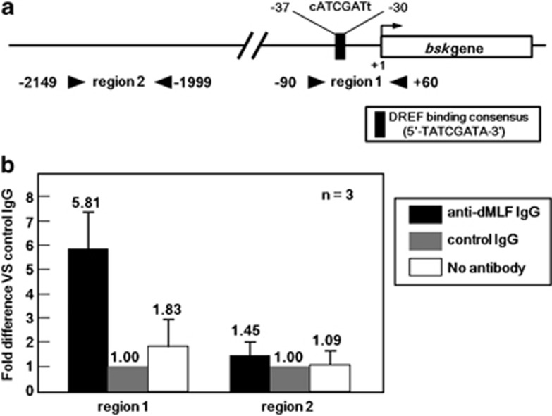
Figure 4.
(a) Schematic illustration of the DREF-binding consensus sequence in the 5′-flanking region of the bsk gene. Arrowheads show the positions of primers used for the ChIP assays for two genomic regions (region 1, proximal, and region 2, distal). (b) Chromatin immunoprecipitation results. The data shown are derived from quantitative real-time PCR analysis of two genomic regions 1 and 2. Chromatin from S2 cells was immunoprecipitated with either anti-dMLF IgG or control rabbit IgG. The fold different values are for anti-dMLF IgG immunoprecipitated samples compared with the corresponding control rabbit IgG immunoprecipitated sample defined as 1.00. A sample without antibody treatment was also included as a negative control (no antibody column).

