
Figure 2.
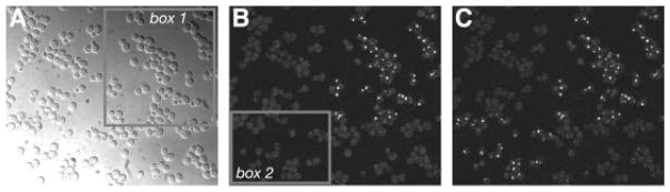
Photoactivation of Erg6p-PA–GFP. Live cells expressing Erg6p tagged at the chromosomal locus with single PA–GFP were examined using a Nikon C1 laser scanning confocal microscope. (A) DIC image. Cells within box 1 were selectively irradiated with a 405 nm laser for 20 s. (B) GFP fluorescence image after irradiation of box 1. Cells within box 2 were subsequently irradiated as in (A). (C) GFP fluorescence image after irradiation of box 2. Note that cells that were photoactivated by the first irradiation remained fluorescent after the second irradiation. Strain: YWL492, Erg6p-PA–GFP