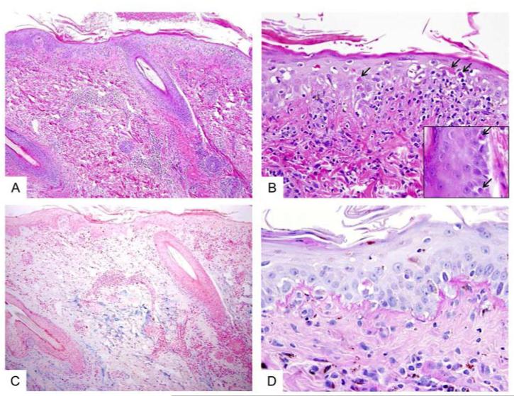
Fig 2. Histopathology of lupus-like lesions.
A, Skin biopsy shows a mild perivascular chronic inflammatory infiltrate in the superficial and deep dermis. The overlying epidermis shows hyperparakeratosis and interface vacuolar changes. (H&E stain, original magnification = 100x). B, Higher magnification highlights frequent necrotic/dyskeratotic keratinocytes (arrows) in the epidermis and along the infundibular portions of hair follicles (inset). (H&E stain, original magnification = 400x; inset = 600x). C, An Alcian Blue stained section shows abundant mucin deposition in the reticular dermis. (Alcian blue stain, original magnification = 100x). D, PAS-D stained section shows irregularly thickened basement membrane along the dermal-epidermal junction. (PAS-D stain, original magnification = 600x)

