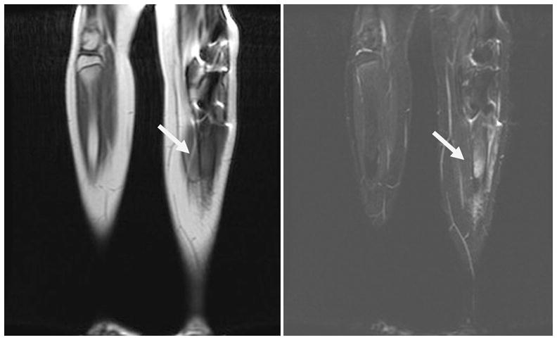
Figure 3.

Twelve year old boy with history of bilateral retinoblastoma as well as previous metastatic disease to the right femur. Whole-body MRI (WB-MRI) showed new signal abnormality within the left tibial shaft on T1-weighted sequences (left) and T2 STIR (right). The patient later reported a several week history of left leg pain. Biopsy revealed a diagnosis of osteosarcoma of the left tibia.
