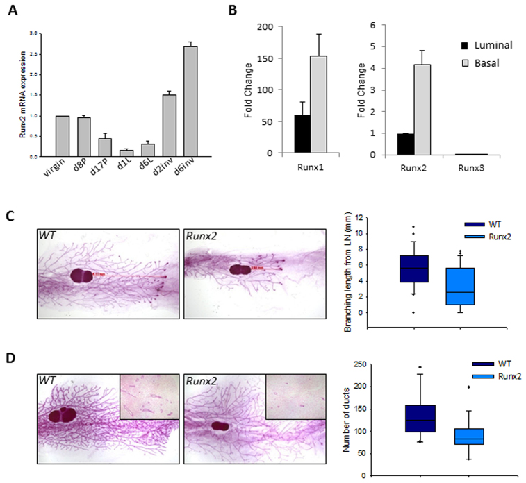
Fig. 2.
Transgenic expression of Runx2 perturbs pubertal mammary development. (A) qRT-PCR of Runx2 throughout murine mammary development. Expression levels relative to wild-type 12-week-old virgins; data are means ± s.d. (P, pregnant; L, lactating; inv, involution, d, day). (B) qRT-PCR of Runx expression in basal/myoepithelial and luminal epithelial cell populations sorted by FACS based on CD29 and CD24 surface markers (see text for details). Runx1 is plotted on a different y-axis owing to the higher levels of expression. Runx3 expression was not detectable in either population. Expression normalised to Gapdh is relative to luminal Runx2; data are means of three independent samples ± s.d. (C) Whole-mounts of 6-week-old mammary glands. Elongation from lymph node (LN) in weight- and litter-matched WT glands is greater than in MMTV-Runx2 glands. Ductal elongation lengths as represented by the red arrows are quantified in the box plot (P=0.002; WT n=28; Runx2 n=23). Dots represent outliers. (D) Whole-mounts of 8-week-old MMTV-Runx2 glands reveal a reduction in tertiary side-branching in Runx2 glands compared with WT glands. Number of ducts per H&E section were counted for each sample (inset images); graph shows a significant reduction in average duct number in Runx2 mammary glands (P=0.01; WT n=15; Runx2 n=16). Dots represent outliers. Whole-mounts, 6.5× magnification; H&Es, 40× magnification.

