
Fig. 6.
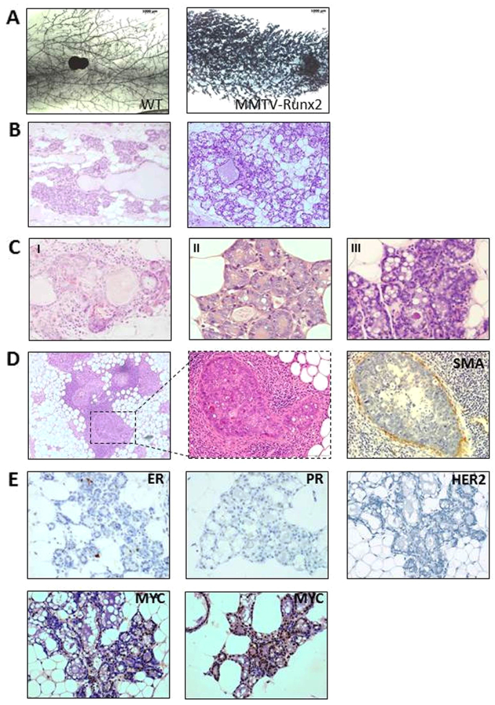
Mammary glands of aged MMTV-Runx2 females display abnormal hyperplastic and pre-neoplastic changes. (A) Representative whole-mounts of aged MMTV-Runx2 and WT littermate controls (8× magnification). (B) Representative images of H&E sections showing diffuse hyperplasia in two independent MMTV-Runx2 transgenic glands with evidence of secretory hyperplastic lesions and dilated ducts (100× magnification). (C) Abnormal features observed in aged MMTV-Runx2 glands, such as distorted acini with lobular fibrosis and chronic inflammatory cell infiltrate (I), and alveolar hyperplasia with luminal cells exhibiting large nuclei and prominent nucleoli (II,III) (images shown at 400× magnification). (D) Ductal carcinoma in situ (DCIS) in an MMTV-Runx2 female; middle panel is higher magnification of boxed area. Smooth muscle actin (SMA) staining shows an intact basal/myoepithelial layer. (E) Hyperplastic lesions are negative for ER, PR and HER2 but show positivity for MYC as determined by immunohistochemistry (400× magnification).