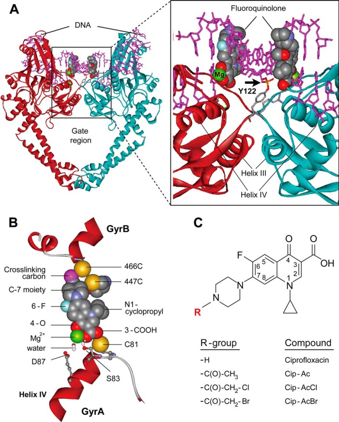
FIGURE 1.
Structures of type II topoisomerase-DNA-quinolone complex and quinolones. A, cleaved complex. The side view of the three-dimensional structure of Acinetobacter baumannii topoisomerase IV bound to DNA and moxifloxacin (Protein Data Bank code 2XKK) is shown in which the DNA gate region is illustrated in an expanded view (right). Moxifloxacin is depicted in a space-filling representation; the arrow indicates the covalent bond between the DNA end and GyrA-Tyr122. One GyrA subunit is shown in maroon, the other is shown in turquoise, and DNA is pink. GyrB is omitted for clarity. B, enlargement of the quinolone-binding region. The proximity of GyrB cysteine substitutions to the cross-linking carbon of Cip-AcCl is shown. Other fluoroquinolone moieties and GyrA amino acids are present to provide orientation. C, structures of quinolones. Ciprofloxacin is shown along with the modified derivatives used in this work.

