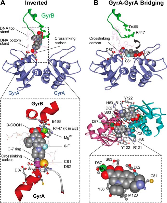
FIGURE 8.

Models for binding of ciprofloxacin to gyrase-DNA complexes with interaction between fluoroquinolone C-7 ring and GyrA81. A, inverted configuration of the published x-ray crystal structure (Protein Data Bank code 2XKK). In the enlargement, GyrB447 is Arg in M. smegmatis and Lys in E. coli. B, GyrA-GyrA bridging model (from Protein Data Bank code 1AB4). The top portion shows one molecule of cross-linked ciprofloxacin in a bridging pocket. The curved arrow indicates the rotation needed to achieve the inverted structure shown in A. The center portion shows two antiparallel pockets filled with fluoroquinolone. The bottom portion shows a detail of nearby GyrA amino acid residues; GyrA87 and GyrA81 are on different GyrA subunits. In both panels, the cross-linking carbon is labeled.
