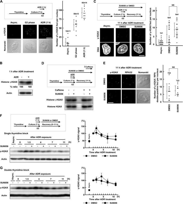
FIGURE 4.
DSB repair is largely unaffected by SFK inhibition. A, HeLa S3 cells were exposed to 110 nm Adriamycin (ADR) in G2 phase as described under “Experimental Procedures” and fixed for immunofluorescence microscopy. After fixation, the cells were stained with anti-γ-H2AX antibody. B, cells were treated as in A. SDS lysates were prepared and probed with the indicated antibodies. C and E, cells were treated as in A and fixed 11 h after the end of Adriamycin exposure. One hour before Adriamycin exposure, 5 μm SU6656 was added. D, the same experiment as in Fig. 3F. Cells were treated as in C and E, and SDS lysates were prepared. One hour before harvest, 5 mm caffeine was added. F and G, HeLa S3 cells were exposed to 110 nm Adriamycin for 1 h from 6 h after release from thymidine block and allowed to recover for the indicated times. SDS lysates were prepared and probed with the indicated antibodies. Cells were synchronized by single thymidine block for 16 h (F) or double thymidine block (G). Each graph represents results obtained from two independent experiments. NS, not significant. Error bars represent S.D. Scale bar, 10 μm in all panels. Async., asynchronously growing cells.

