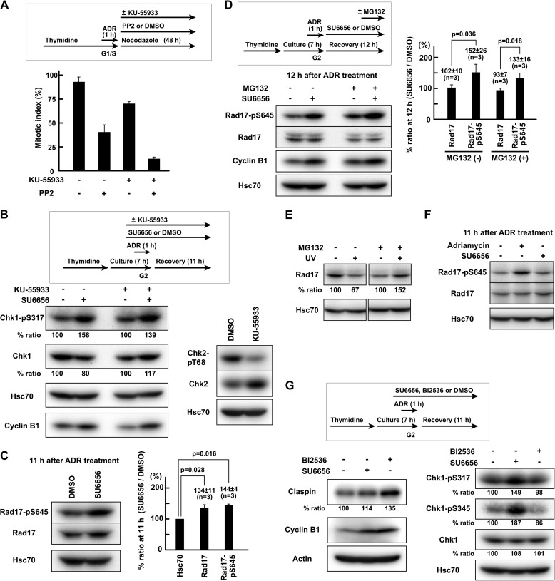
FIGURE 5.
SFKs regulate Rad17 phosphorylation independently of proteasomal degradation of Rad17. A, HeLa S3 cells were treated as in Fig. 1B except that 20 μm PP2 and/or 10 μm KU-55933 was used. The mitotic index was determined after 48 h of recovery. B, cells were exposed to 110 nm Adriamycin (ADR) in G2 phase as described under “Experimental Procedures.” One hour before Adriamycin exposure, 10 μm KU-55933 was added. SDS lysates were prepared. C, the same experiment as in Fig. 3, B–E. Cells were exposed to 110 nm Adriamycin in G2 phase as described under “Experimental Procedures,” and SDS lysates were prepared 11 h after the end of Adriamycin exposure. D, HeLa S3 cells were treated as in C except that 5 μm SU6656 was added after Adriamycin exposure. Two hours before harvest, 10 μm MG132 was added. E, HeLa S3 cells were UV-irradiated (10 J/m2), allowed to recover for 4 h, and solubilized in SDS-PAGE sample buffer. One hour before UV irradiation, 10 μm MG132 was added. The data were obtained from the same blot. F, the same experiment as in C. G, HeLa S3 cells were treated as in C. Cells were exposed to SU6656 or BI2536. p values were calculated using t test. Error bars represent S.D.

