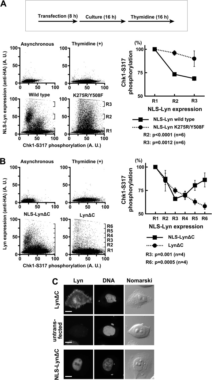
FIGURE 8.
SFKs promote checkpoint silencing through nuclear protein tyrosine phosphorylation. A and B, HeLa S3 cells were transfected and subsequently exposed to thymidine for 16 h. The cells were fixed, stained with the indicated antibodies, and analyzed by flow cytometry. The intensity of the Chk1-Ser(P)317 signal was quantitated in each region (Rn) and expressed relative to that obtained with untransfected cells (R1). p values were calculated using t test. Error bars represent S.D. C, the cells treated as in B were fixed for immunofluorescence and stained as indicated. An untransfected cell on the same coverslip as LynΔC-transfected cells is shown as a control. Scale bar, 10 μm in all panels. A. U., arbitrary units.

