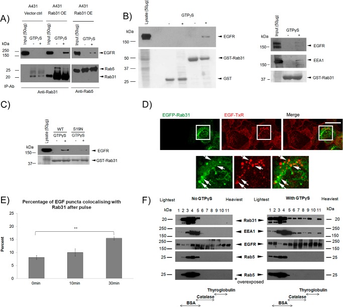
FIGURE 4.
Rab31 associates with EGFR in a high molecular weight complex that includes its effector EEA1 but not Rab5. A, EGFR was coimmunoprecipitated (IP) with Rab31 or Rab5 antibody (ab), respectively, using 1 mg of lysates from cells transfected with vector alone (Vector ctrl) and Rab31 (Rab31 OE), respectively, with or without GTPγS loading. B, 1 mg of A431 cell lysate (left panel) or HeLa cell lysate (right panel) with and without GTPγS was incubated with 20 μg of GST or GST-Rab31 and glutathione beads, and the ability of the GST fusion proteins to pull down EGFR was analyzed by Western blot analysis. The GST fusion proteins were visualized with Ponceau S stain. C, 1 mg of A431 cell lysate with and without GTPγS was incubated with 20 μg of GST-Rab31 or GST-Rab31S19N (S19N) and glutathione beads, and the ability of the GST fusion proteins to pull down EGFR was analyzed by Western blot analysis. The GST proteins were visualized with Ponceau S stain. D, A431 cells stably transfected with EGFP-tagged Rab31 (EGFP-Rab31) were pulsed with 0.5 μg/ml EGF-TxR, fixed after 30 min, and analyzed for colocalization between EGFP-Rab31 (green) and EGF-TxR (red). Bottom panel, the boxed areas in the upper panel, enlarged ×2. Arrows indicate structures positive for both EGFP-Rab31 and EGF-TxR. Scale bar = 20 μm. E, the percentage of EGF-TxR-positive puncta that are also positive for EGFP-Rab31 was quantified from cells fixed after a 0-, 10-, and 30-min chase and graphically represented as a percentage of total EGF-TxR puncta counted. 34 cells in three independent experiments were analyzed, and data are shown as mean ± S.E. **, p < 0.01; Student's t test. F, 2 mg of A431 Rab31 OE lysates with and without GTPγS loading was resolved by glycerol gradient sedimentation. Fractions were collected, and TCA was precipitated and analyzed by Western blot analysis for Rab31, EGFR, Rab5, and EEA1 (an effector for both Rab5 and Rab31). The membrane blot for Rab5 was also overexposed. The position in the gradient that contains the molecular size markers BSA (67 kDa), catalase (240 kDa), and thyroglobulin (660 kDa) is indicated.

