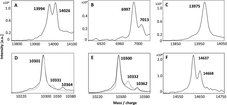
FIGURE 4.

MALDI-TOF spectra of persulfurated Rhd_2599, TusA, and DsrC proteins. 30 μm protein solutions were incubated with a suitable sulfur substrate or sulfur-donating protein for 60 min at 30 °C. A, Rhd_2599 (mass 13,991 Da) after incubation with 2 mm thiosulfate. B, Rhd_2599 after incubation with 0.5 mm GSSH. C, the Rhd_2599-Cys-64Ser variant protein (mass 13,975 Da) is shown after incubation with 2 mm thiosulfate. D, TusA (mass 10,300 Da) after incubation with 2 mm sulfide. E, TusA after incubation with Rhd_2599 and thiosulfate. In D and E, two superimposed spectra obtained for two individually prepared samples are shown. F, persulfidic TusA was incubated with 30 μm DsrC (mass 14638 Da) as acceptor molecule. Binding of sulfur atoms is indicated by an additional mass of 32 Da. Note that in B the spectrum for the double-charged protein is shown. a.u., absorbance units.
