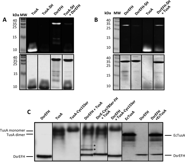
FIGURE 6.
Sulfur transfer between and interaction of TusA and DsrEFH. In panel A the capacity of TusA for sulfur transfer to DsrEFH was analyzed by the 1,5-IAEDANS method. In panel B the results for the reverse reaction, i.e. sulfur transfer from persulfurated DsrEFH to TusA, are presented. In both cases (A and B), the respective persulfurated donor protein was incubated with the acceptor protein for 60 min at 30 °C before the reaction mixtures were treated with 1,5-IAEDANS and DTT as described under “Experimental Procedures.” Samples were then electrophoresed on non-reducing 15% SDS-polyacrylamide gels. Gels were analyzed under UV-light (upper gels of panel A and B) and subsequently stained with Coomassie Brilliant Blue (lower gels of panels A and B). Part C shows the interaction of DsrEFH and TusA from A. vinosum (TusA) and E. coli (EcTusA), respectively. For the interaction experiments, 800 pmol of TusA/EcTusA were incubated with 100 pmol of DsrEFH (wild type and mutant proteins). Samples were then analyzed via native SDS-PAGE (7.5%). Bands arising from interaction complexes are marked with stars. Molecular mass (MW) of marker proteins is given in kDa.

