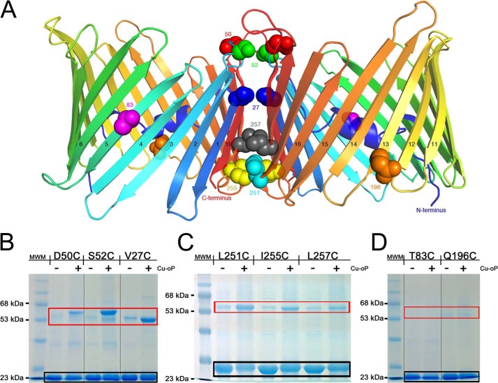
FIGURE 8.
Cross-linking experiments validate the dimer interface. A, cartoon representation of the zfVDAC2 putative dimer with sites of cysteine mutations displayed in spheres, differently colored by position. B, 12% SDS-PAGE for zfVDAC2 single-cysteine mutants close to the cytosolic side. C, 12% SDS-PAGE for zfVDAC2 single-cysteine mutants close to the IMS. D, 12% SDS-PAGE of non-cross-linking single-cysteine mutants at positions outside of the putative dimer interface. Black box, monomeric zfVDAC2; red box, dimeric forms. MWM, molecular weight markers. Cu-oP, dichloro(1,10-phenanthroline)Cu(II) Groupings of images from different parts of the same gels are marked by vertical lines.

