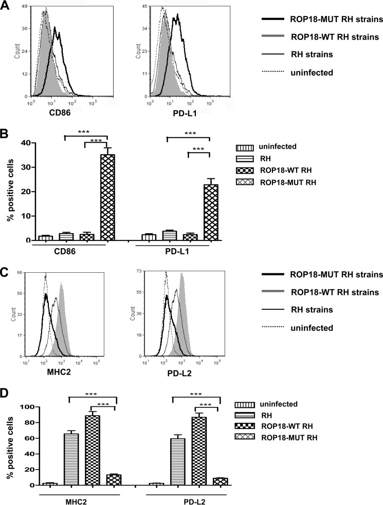
FIGURE 8.
Inducing the M2 phenotype with type I strains expressing wild-type ROP18. A, U937 cells were infected with ROP18-WT RH strain, ROP18-MUT RH strain, or RH strain. At 24 h post-infection, macrophages were stained for CD86 or PD-L1, and fluorescence-activated cell sorting assays were performed (dotted lines, uninfected macrophages; solid lines, macrophages infected with RH strain; heavy lines, macrophages infected with ROP18-MUT RH strain; shaded, macrophages infected ROP18-WT RH strains). B, histograms depict the percentage of positively stained CD86 or PD-L1 cells analyzed in A. Indicated values are the means ± S.D. of triplicates. ***, p < 0.001, compared with the RH strain control. C, U937 cells were infected with ROP18-WT RH strain, ROP18-MUT RH strain, or RH strain. At 24 h post-infection, macrophages were stained for MHC2 or PD-L2, and fluorescence-activated cell sorting assays were performed (dotted lines, uninfected macrophages; solid lines, macrophages infected with RH strain; heavy lines, macrophages infected with ROP18-MUT RH strain; shaded, macrophages infected ROP18-WT RH strain). D, the histograms depict the percentage of positively stained MHC2 or PD-L2 cells analyzed in C. Indicated values are the means ± S.D. of triplicates. ***, p < 0.001, compared with the RH strain control.

