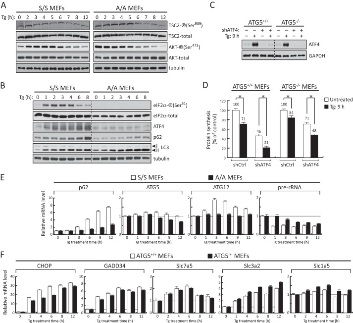
FIGURE 4.
Induction of ATF4 during prolonged ER stress contributes positively to protein synthesis rates independently of autophagy. A and B, Western blot analysis of extracts from S/S and A/A cells treated with Tg (400 nm) for the indicated times. C, Western blot analysis of extracts from ATG5+/+ and ATG5−/− MEFs infected with adenoviral particles expressing either control shRNA or shRNA against ATF4. D, protein synthesis was measured by [35S]Met/Cys incorporation into proteins in either untreated or Tg-treated ATG5+/+ and ATG5−/− MEFs following 3 days of adenovirus infection for the corresponding shRNAs. Data obtained were normalized to the values of the respective cells expressing control shRNA without Tg treatment. Results are the mean of triplicate determinations. The asterisks indicate p < 0.05. E and F, RT-qPCR analysis of mRNAs isolated from S/S, A/A, ATG5+/+, and ATG5−/− MEFs treated with Tg for the indicated times. Data were normalized to GAPDH values and expressed as a ratio to the value of untreated samples. Error bars represent S.E.

