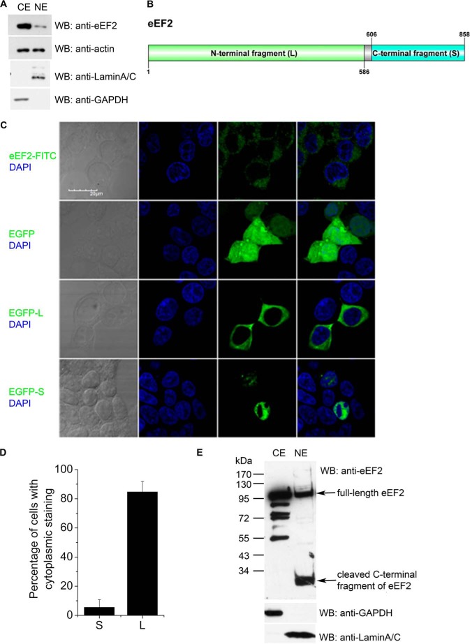
FIGURE 2.
Nuclear localization of full-length eEF2 and its small cleaved fragment. A, eEF2 could translocate into nucleus. HEK293T cells were fractionated and blotted with indicated antibodies. CE, cytosolic fraction; NE, nuclear fraction. Anti-C-terminal eEF2 showed the different distribution of eEF2 in cytosolic and nuclear fractions. Anti-actin antibody showed similar amounts of cytosolic and nuclear proteins. Lamin A/C and GAPDH were used as nuclear and cytosolic markers, respectively. WB, Western blot. B, diagram of the eEF2 plasmid constructs. Large and small fragments of eEF2 were constructed as indicated. This diagram was drawn by Group-based prediction System (DOG 2.0). C, HEK293T cells were stained with anti-N-terminal eEF2 antibody (top row, green color) or transfected with EGFP control plasmid, EGFP-L, and EGFP-S plasmids. The images were taken with confocal microscopy (GFP, green color). Nuclei were stained with DAPI (blue color). D, quantification of data in C. Results are mean ± S.D. of at least 50 transfected cells. E, cleaved small fragment of eEF2 accumulated in nucleus. HEK293T cells were fractionated. Anti-C-terminal eEF2 antibody showed the cleaved eEF2 in nucleus. GAPDH was used as a cytosolic marker, and lamin A/C was used as nuclear marker. Data are representatives of three independent experiments.

