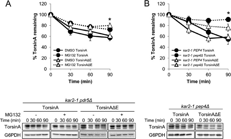
FIGURE 6.
Loss of Kar2/BiP function causes torsinA and TorsinAΔE to be degraded by the proteasome and by the vacuole. CHX chases were performed as described before (see legend for Fig. 3). A, torsinA and torsinAΔE degradation in the kar2-1 pdr5Δ strain treated with DMSO or 100 μm MG132 for 30 min before addition of CHX (*, p < 0.02 for DMSO torsinA versus MG132 torsinA treatment; p < 0.02 for DMSO torsinAΔE versus MG132 torsinAΔE treatment). B, torsinA and torsinAΔE degradation in the kar2-1 pep4Δ strain. To facilitate the comparison, we included in the graph the data from the kar2-1 PEP4 strain from Fig. 3A (*, p < 0.002 for kar2-1 PEP4 torsinA versus kar2-1 pep4Δ torsinA; p < 0.02 for kar2-1 PEP4 torsinAΔE versus kar2-1 pep4Δ torsinAΔE). Experiments were performed at least twice, with ≥2 independent replicates per experiment. Representative Western blots are shown below.

