
FIGURE 12.
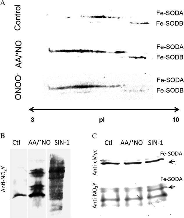
Cellular detection of Fe-SODA nitroxidative modifications. A, two-dimensional gel electrophoresis. T. cruzi Fe-SODA overexpressers were treated with AA (5 μm) plus NOC-12 (5 mm; t½ = 100 min at pH 7.4), SIN-1 (5 mm), or ONOO− (300 μm) for 3 h at room temperature. After treatment, samples were processed as described under “Experimental Procedures” and subjected to two-dimensional electrophoresis. Membranes were probed with the specific Fe-SODA and Fe-SODB antibodies. B, 3-nitrotyrosine detection in parasites. Following treatment in the conditions described in A, parasite extracts were separated in 15% SDS gels and transferred to nitrocellulose membranes. Membranes were probed with the specific anti-3-nitrotyrosine antibody. C, immunoprecipitation of nitrated Fe-SODA. Parasite extracts as above were incubated overnight at 4 °C in the presence of the monoclonal c-Myc antibody that recognized the 9E10 epitope of Fe-SODA in the presence of protein A/G-agarose as described under “Experimental Procedures.” Immunoprecipitated proteins were run in 15% SDS-gel electrophoresis, electrotransferred to nitrocellulose, and revealed using anti-c-Myc antibody and anti-3-nitrotyrosine antibody and as described under “Experimental Procedures.”