
FIGURE 5.
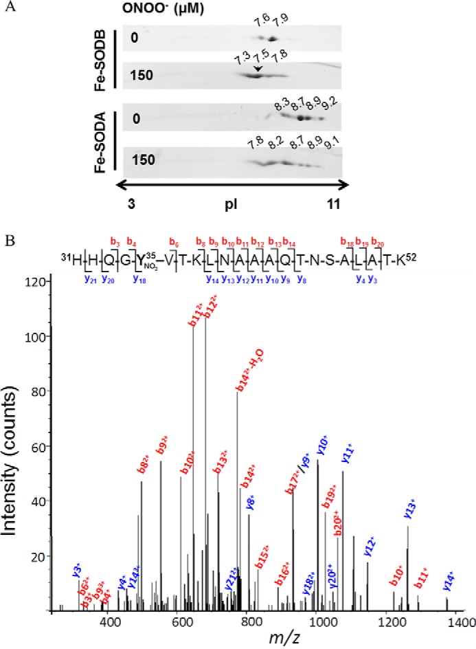
Peptide mapping of T. cruzi Fe-SODs after peroxynitrite treatment. A, T. cruzi Fe-SODs (8 μm) were treated with peroxynitrite (150 μm) in sodium phosphate buffer (0.2 m) at pH 7.4 and 25 °C. Two-dimensional gel electrophoresis was performed as described under “Experimental Procedures.” The arrowhead shows the selected Fe-SODB spot of pI 7.5 analyzed by mass spectrometry. B, MS/MS spectrum of triply charged ion at m/z 790.6 (MH+ 2369.7, retention time = 30.3 min) from a tryptic digestion of peroxynitrite-treated Fe-SODB spot (pI = 7.5, indicated with an arrow in A). The major N-terminal (b, red-labeled) and C-terminal (y, blue-labeled) fragment ions that allowed the sequence 31–52 assignment that includes a nitrated tyrosine residue (mascot ion score = 61; p < 0.05) are shown. Inset, amino acid sequence of peptide 31–52, indicating major b and y ions detected by full-scan MS/MS.