
FIGURE 7.
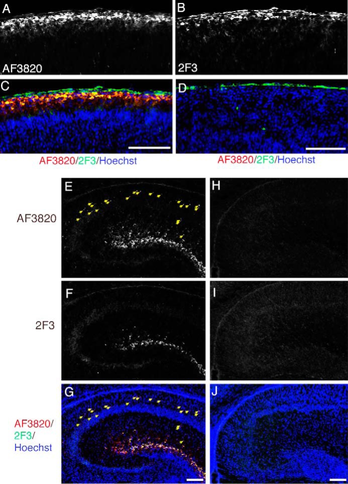
Localization of full-length and cleaved Reelin. Frozen sections from the cerebral cortices of postnatal day 1 (P1) wild-type (A–C) or reeler (D) mice were immunostained with AF3820 (A) and mAb 2F3 (B). C, merged image of A (red), B (green), and Hoechst33342 nucleus staining (blue). D, merged image of signals from AF3820 (red), mAb 2F3 (green), and Hoechst33342 (blue) staining. The pia mater gave a background staining when mAb 2F3 was used (the green signals outside of the brain in C and D). E–J, frozen sections of hippocampi of P1 wild-type (E–G) or reeler (H–J) mice were coimmunostained with AF3820 (E and H) and mAb 2F3 (F and I). The merged images with Hoechst33342 nucleus staining (blue) are shown in G and J. The yellow arrows indicate punctate signals that are positive for AF3820 but not 2F3. Scale bars = 100 μm.