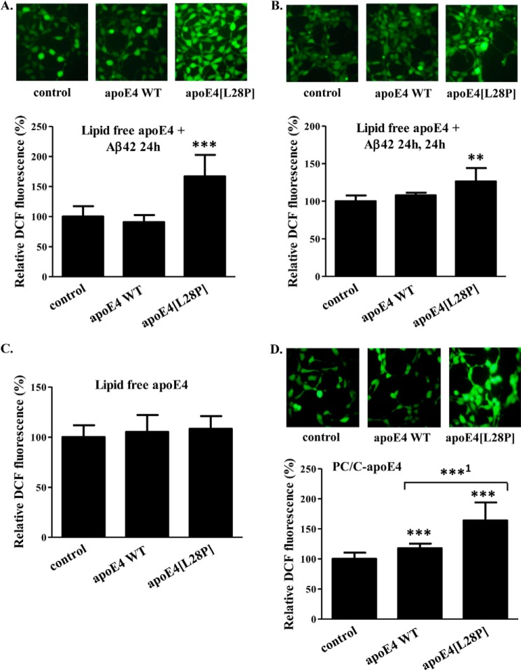
FIGURE 11.
Effect of lipid free or lipoprotein particle-associated WT apoE4 and apoE4[L28P] in the presence or absence of Aβ42 on ROS formation by SK-N-SH cells. A, SK-N-SH cells were incubated with 25 ng/ml Aβ42 in the absence (control) or presence of 375 nm lipid-free WT apoE4 or apoE4[L28P] for 24 h. B, SK-N-SH cells were incubated with 25 ng/ml Aβ42 in the absence (control) or presence of 375 nm lipid-free WT apoE4 or apoE4[L28P] for 24 h and then washed and incubated in fresh medium without Aβ42 and apoE4 forms for another 24 h. C, SK-N-SH cells were incubated in the absence (control) or presence of 375 nm lipid-free WT apoE4 or apoE4[L28P] for 24 h (without Aβ42). D, SK-N-SH cells were incubated in the absence (control) or presence of 375 nm lipoprotein particle-associated (PC/C) WT apoE4 or apoE4[L28P] for 24 h (without Aβ42). At the end of each incubation period, the cells were incubated with 2′,7′-dichlorofluorescein diacetate for 45 min. The formation of ROS was measured by detection of fluorescent DCF emitted from cells using a fluorescence microscope, as described under “Experimental Procedures.” The DCF fluorescence of cells incubated with lipid-free or lipoprotein particle-associated apoE4 forms in the presence or absence of Aβ42 is shown relative to DCF fluorescence of control cells set as 100%. DCF fluorescence intensity was measured for at least 40 cells from the fluorescent images of each sample, as described under “Experimental Procedures,” and the relative fluorescent intensity was taken as the average of the values of at least five images for each experiment. Values are the means ± S.D. (n = 20) of four experiments. **, p = 0.053 versus control; ***, p < 0.0001 versus control; ***1, p = 0.0001 versus WT.

