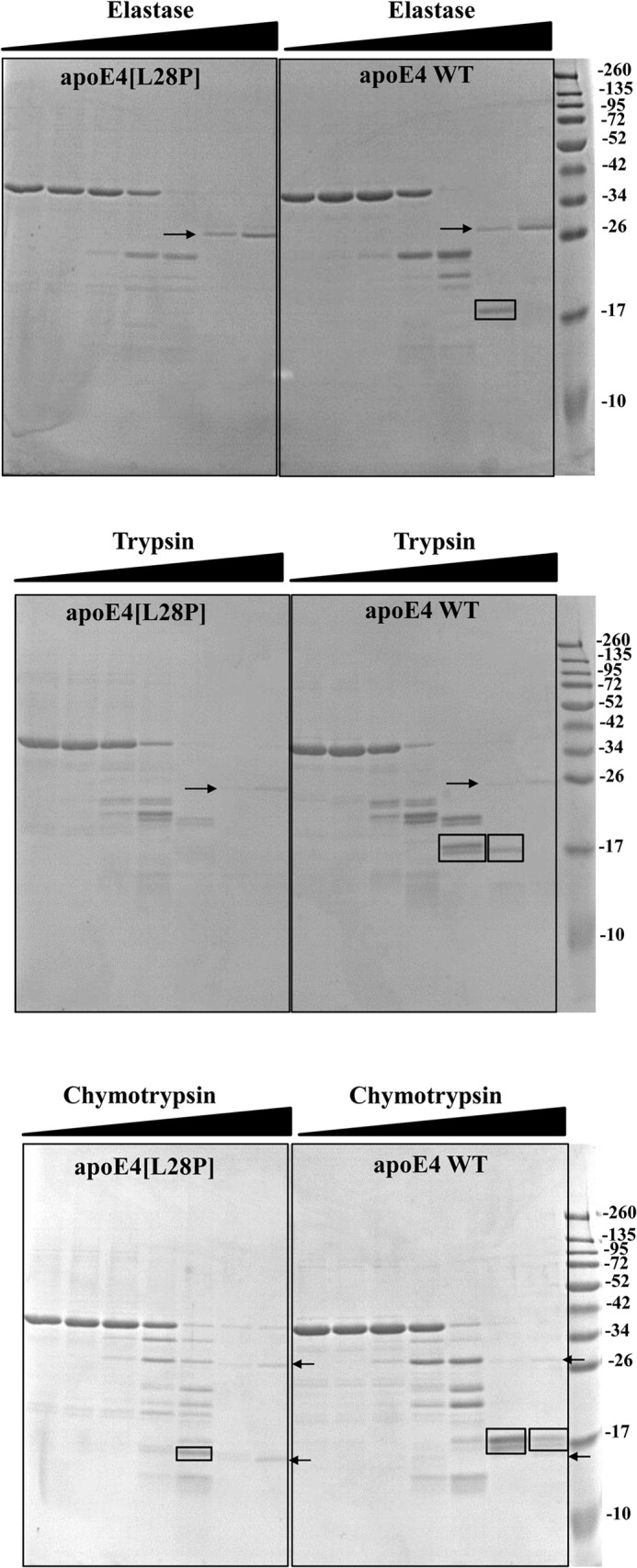
FIGURE 3.

Protease digestion sensitivity of WT apoE4 and apoE4[L28P]. WT and mutant apoE4 were incubated for 1 h at room temperature with increasing amounts of elastase (top panel), trypsin (center panel), and chymotrypsin (bottom panel) as described under “Experimental Procedures.” Reactions were stopped by the addition of PMSF and analyzed on SDS-PAGE. Arrows indicate the bands that correspond to the protease (only visible for the highest concentrations used). Rectangles highlight different apoE4 fragments that accumulate in WT apoE4 but are degraded in the mutant apoE4 or apoE4 fragments that are observed for chymotrypsin proteolysis of mutant apoE4 but not of WT apoE4.
