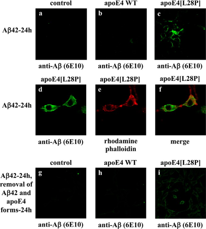
FIGURE 8.

Fluorescence confocal laser-scanning microscopy of SK-N-SH cells incubated in the presence of Aβ42 and WT apoE4 or apoE4[L28P]. SK-N-SH cells were incubated with 25 ng/ml Aβ42 in the absence (control) or presence of 375 nm lipid-free WT apoE4 or apoE4[L28P] for 24 h, as indicated (a–f). SK-N-SH cells were incubated with 25 ng/ml Aβ42 in the absence (control) or presence of 375 nm lipid-free WT apoE4 or apoE4[L28P] for 24 h and then washed and incubated in fresh medium without Aβ42 and apoE4 forms for another 24 h, as indicated (g–i). Aβ immunostaining of cells was detected with the antibody 6E10, followed by an FITC-conjugated secondary antibody (a–d, g–i, green). F-actin was stained with rhodamine phalloidin (e, red). The merger of images d and e is shown in f.
