
Figure 2.
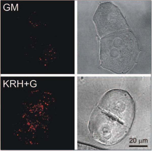
Confocal and wide field microscopy of LysoTracker Red (LTR) uptake in hepatocytes after autophagic induction. Cultured hepatocytes were loaded with LTR (200 nM, 20 min) and incubated for 70 min in complete growth medium (GM, upper panels) or in KRH plus 1 μM glucagon (KRH+G, lower panels). The left panels show representative superimposed through-focus confocal images of red LTR fluorescence of hepatocytes in growth medium (upper left panel) or KRH plus glucagon (lower left panel). The right panels are corresponding bright images.