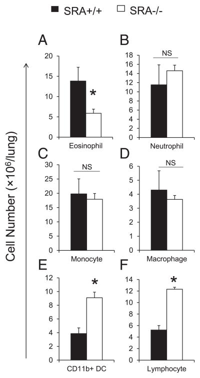
FIGURE 3.
Effect of SRA on the accumulation of pulmonary leukocytes. Lung leukocytes were isolated from infected SRA+/+ and SRA−/− mice at 3 wpi, Ab stained, and analyzed using flow cytometry as per Materials and Methods. In brief, leukocytes were identified as CD45+ cells. Next, additional gates were set up to identify lymphocytes (CD3+ and CD19+), neutrophils (Ly6G+/CD11b+), eosinophils (SSChigh/CD11cint), Ly6C+ monocytes (CD11c−/Ly6C+/CD11b+), macrophages (autofluorescence+/CD11c+), and CD11b+ DCs (autofluorescence−/CD11c+/CD11b+). The total number of CD45+ leukocytes was determined by multiplying the frequency of CD45+ by the total number cells. Total numbers of eosinophils (A), neutrophils (B), monocyte (C), macrophages (D), CD11b+ DCs (E), and lymphocytes (F) were determined by multiplying the frequency of each subset by the total number of CD45+ leukocytes at 3 wpi. Data represent mean ± SEM pooled from one separate experiments; n ≥ 6 for each of the analyzed parameters; *p <0.05 in comparison between SRA+/+ and SRA−/− mice. NS, No significant difference between SRA+/+ and SRA−/− mice.

