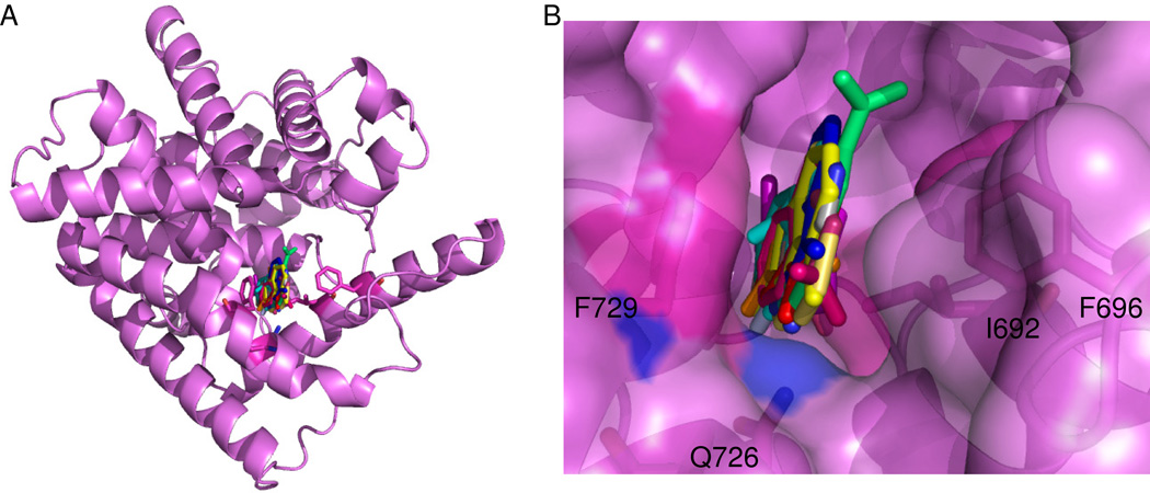
Figure 2.
(A) Location of fragments binding to PDE10A in this study. All fragments were found at the primary binding pocket typically occupied by cAMP or cGMP14. Fragments were not located at any other binding site on the protein. (B) Close-up of a superposition of all co-crystal structures of fragments bound in the primary binding pocket of PDE10A. Fragments are found along a single plane and oriented between F729 and I692, F696. Q726 is making at least one hydrogen bond with all of the fragment hits.

