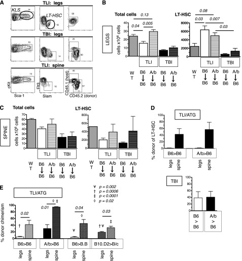
Figure 4.
Engraftment of HSC differs in distinct marrow sites after TLI/ATG. Thy1.2.CD45.1 B6 mice underwent TLI/ATG or sublethal TBI (475 cGy) and were injected IV with 10 000 congenic Thy1.1.CD45.2 B6 or miAg-mismatched AKR/b (Thy1.1. CD45.2) KTLS-HSC. At 2 weeks (A-D) and 3 months (E) post-HCT, the level of donor chimerism of HSC in the BM of the legs vs the vertebrae was assessed. (A) Representative FACS plots and gating schema reveals no evidence of engrafted donor LT-HSC in the legs after TLI/ATG (top), and mixed chimerism after TBI (middle). After TLI/ATG, donor and host LT-HSC were detectable in the BM of irradiated spine (bottom). (B) Compiled data on absolute cell counts per 2 legs in WT mice (dark gray); TLI/ATG-treated B6 recipients of B6-HSC (solid white); AKR/B (A/b)-HSC (white with stripes); TBI-conditioned B6 recipients of B6-HSC (solid black); and AKR/B-HSC (black with stripes). AKR/b grafts resulted in higher cell counts in B6 recipients compared with B6 grafts (left). Absolute numbers of LT-HSC per 2 legs in WT mice; TLI/ATG-treated B6 recipients of B6-HSC and AKR/B-HSC; and TBI-conditioned B6 recipients of B6-HSC and AKR/b-HSC (right). (C) Compiled absolute cell counts per 5 vertebrae from spines of WT mice, TLI/ATG vs TBI-treated B6 recipients of B6-HSC and AKR/b-HSC, respectively, revealing insignificant differences between groups (left). Absolute numbers of LT-HSC per 5 vertebrae of WT mice, TLI/ATG vs TBI-treated B6 recipients of B6-HSC and AKR/b-HSC showing no significant differences (right). (D) Compiled data on percent donor chimerism in TLI/ATG-treated (top) and TBI-treated (bottom) B6 recipients of B6-HSC or AKR/B-HSC in legs and spine, respectively. (E) Percent donor chimerism at 3 months post-HCT in TLI/ATG-treated B6 (H2b) recipients of B6-HSC (H2b), B6 recipients of AKR/b-HSC (H-2b), BALB.B (B.B; H-2b) recipients of B6 HSC, and BALB/c (B/c; H-2d) recipients of B10.D2 HSC (H-2b) in legs (solid) and spines (striped pattern), respectively. N = 3 to 6 animals per experimental group.

