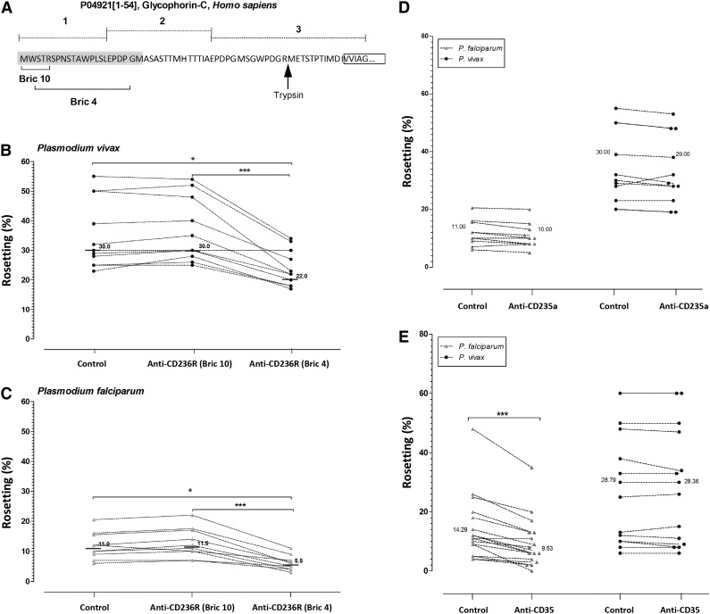
Figure 5.
Antibody blocking rosette formation. (A) Schematic diagram of the target sites of anti-CD236R antibody clone BRIC 10 and anti-CD236R antibody clone BRIC 4 on human CD236R structure. The target site of trypsin on this sialoglycoprotein is shown by the black arrow. (B-C) Rosetting inhibition in P vivax and P falciparum caused by Fab fragments specifically targeting the BRIC 10 and BRIC 4 locations on CD236R. BRIC 4 showed a significant reduction in rosetting of P falciparum isolates (P < .0001) and P vivax isolates (P < .0001) studied. A paired Student t test was conducted. (D) Rosetting inhibition by mouse anti-human CD35 antibody. Unlike in P vivax, this antibody significantly reduced rosetting rate of P falciparum isolates tested (P < .0001). (E) Comparison of rosetting rates between the control and cells incubated with Fab fragments of mouse anti-human glycophorin A antibody from P falciparum and P vivax isolates recruited. There was no significant difference between the control group and the “anti-glycophorin A” group in P vivax and P falciparum isolates studied.

