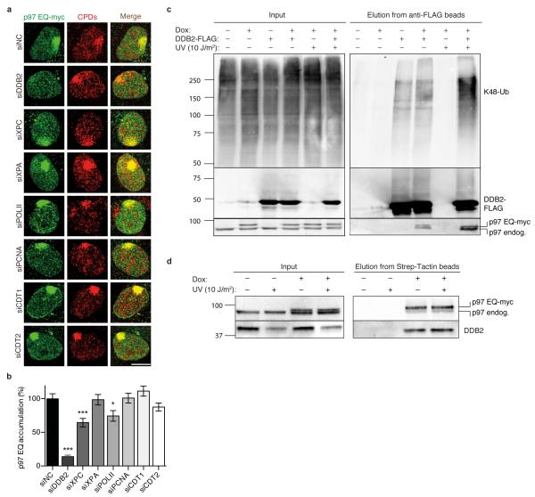
Fig. 2. Recruitment of p97 to UV lesions requires DDB2.
(a) Co-localisation of p97 EQ with CPDs in human cells treated with siRNA targeting the indicated proteins. Samples were probed 15 min after UV irradiation through micropore filters; scale bar, 10 μm. (b) Quantification of p97 EQ accumulation at CPDs as illustrated in Fig. 2a. Error bars, s.e.m. (>300 nuclei from three experiments); *P<0.05, ***P<0.001 relative to siNC control (unpaired two-tailed t-test). (c) UV-stimulated interaction of p97 with a DDB2-containing complex. HEK293 cells were transfected to produce DDB2-FLAG and doxycycline-treated (Dox) for mild p97 EQ expression. DDB2-FLAG was pulled-down from cell lysates using beads coated with anti-FLAG antibodies and eluates were analysed by immunoblotting against K48-ubiquitin, FLAG epitope and p97. Myc-tagged p97 migrates slower than the endogenous counterpart. (d) Interaction of DDB2 with the p97 complex. HEK293 cells were doxycycline-treated (Dox) for mild p97 EQ expression and p97 EQ was pulled-down from cell lysates using beads coated with Strep-Tactin. The resulting protein mixture was analysed by immunoblotting against p97 and DDB2.

