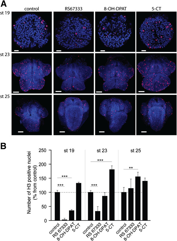
Figure 6.
Effects of different 5-HT receptor agonists on cell proliferation during H. trivolvis development. (A) Representative confocal images of the whole-mount H. trivolvis embryos co-stained with DAPI (blue) and the antibody against phospho-H3S28 (red) after incubation with RS67333, 8-OH-DPAT and 5-CT (5 μM each). Staining with anti-phospho-H3S28 antibody allows visualization of dividing cells. Scale bars are 20 μM for the stage 19, and 50 μM for the stages 23 and 25. (B) The relative number of dividing cells at stages 19, 23 and 25 after 10 h of incubation with RS67333, 8-OH-DPAT and 5-CT. Each value represents the mean ± s.e.m. (n = 10). A statistically significant difference between values is noted (** p <0.01, *** p <0.001).

