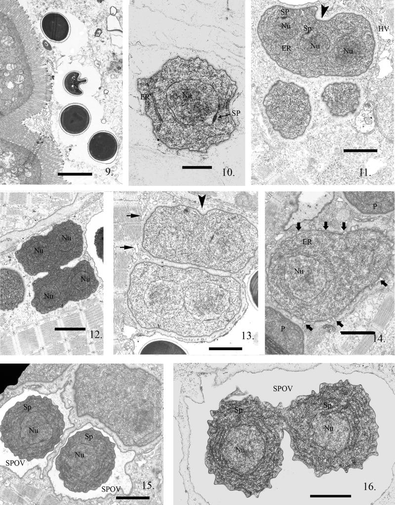
Fig. 9--16.
Electron micrographs (EM) of zebrafish infected with Pseudoloma neurophilia. Fig. 9. Four spores in the intestinal lumen of a larval zebrafish 3 h PE. Scale bar = 2.3 μm. Fig. 10. A uninucleate sporoplasm in the host cell cytoplasm. The nuclear membrane has a dense spindle plaque (SP) indicating this parasite cell is about to undergo karyokinesis. Note the prominent nucleus (Nu), the presence of endoplasmic reticulum (ER) and ribosomes in the sporoplasm. Scale bar = 800 nm. Fig. 11. Proliferative stages in direct contact with the host cell cytoplasm, one containing three nuclei (Nu). Two of the nuclei have prominent SP on their nuclear membranes. The parasite plasmalemma appears to have a glycocalyx-like surface, giving the cells the appearance of a halo surrounding them. Note that karyokinesis is not linked to cytokinesis (arrowhead), resulting in production of multinucleated parasite cells. The host cytoplasm has sarcoplasmic reticulum vacuoles (HV) in the region of the infection. Scale bar = 1.7 μm. Fig. 12. A proliferative cell with four visible nuclei (Nu) in the process of dividing into two multinucleate cells. They will divide repeatedly, resulting in the production of several uninucleate cells. These dividing cells maintain their glycocalyx- like coat, outlining the parasite plasmalemma, during division. Note the presence of small vesicles of the sarcoplasmic reticulum outlining the parasite cells but absent in the glycocalyx-like halo. Scale bar = 2.3 μm. Fig. 13. Two recently divided proliferative cells, each containing two nuclei. The start of a cytoplasmic invagination (arrowhead) indicates this cell is beginning another cytokinesis. Note the presence of host vesicles (short arrows) of sarcoplasmic reticulum abutting the contractile fibers but limited from parasite plasmalemmal contact by the glycocalyx-like coating. Scale bar = 1.4 μm. Fig. 14. Parasite cell starting the transition from proliferative to sporogonic development. Note the difference between it and the abutting proliferative cell (P) membrane which is still covered with its glycocalyx-like surface coat. The plasmalemma has numerous “blisters” starting to emerge (bold arrows) from it and the new plasmalemma is now “thickened” in the areas under the blisters. The blistering continues until it forms an isolation chamber, an SPOV indicating the commencement of sporogony. Scale bar = 1.7 μm. Fig. 15. Two uninucleate (Nu) sporonts (Sp) each in an SPOV. The outer edge of the SPOVs are in contact with the host muscle cell cytoplasm while the sporonts inside them now have a thickened plasmalemma and are isolated from the host cytoplasm. Scale bar = 1.4 μm. Fig. 16. SPOV containing uninucleate (Nu) sporonts (Sp) undergoing cytokinesis. These sporont cells are still narrowly connected and have a thickened plasmalemma and a marked increase in endoplasmic reticulum. Scale bar = 1.2 μm.

