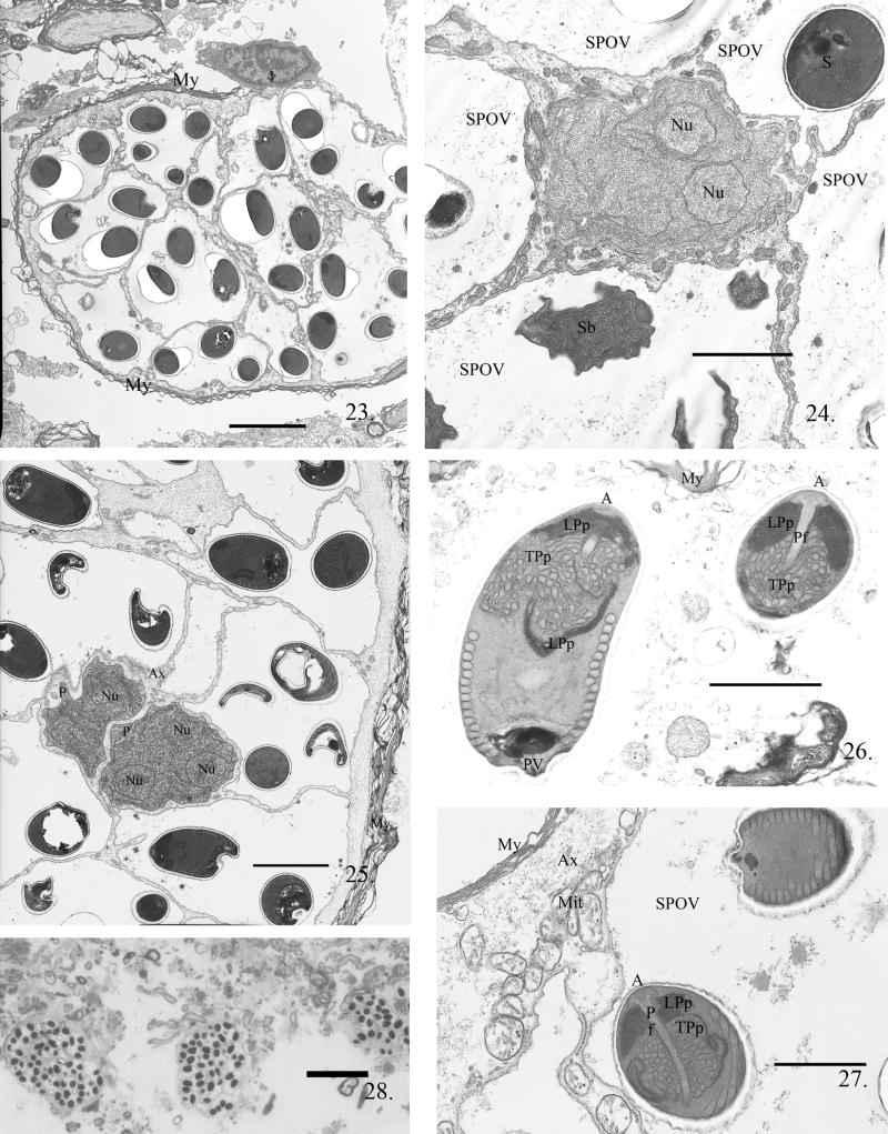
Fig. 23--28.
The myelinated nerves of adult zebrafish infected with P. neurophilia. Fig. 23. Low power image of a myelinated nerve with a long-term infection. The axoplasm filled with 13 SPOVs containing spores; note the presence of a scant amount of axoplasm just inside the myelin (My) layers. Scale bar = 5 μm. The two proliferated cells (P) surround by five SPOVs containing spores (S) and sporoblast (Sb). Fig. 24. Multinucleated proliferative cell in direct contact with remaining axoplasm and surrounded by numerous SPOVs containing spores and one SPOV containing sporoblasts (Sb). Scale bar = 2.3 μm. Fig. 25. Two multinucleate dividing proliferative cells (P) in nerve fiber axoplasm surrounded by SPOVs containing multiple spores. This section is through the edge of the nerve fiber and both the remaining axoplasm (Ax) and myelin (My) are visible. Scale bar = 3 μm. Fig. 26. Two spores in an SPOV. The anterior structure of the spore is well-illustrated in the spores in this section. In the longitudinally sectioned spore, the lamellar polaroplast (LPp) surrounds the tubular portion (TPp) and clearly extends about half the length of the spore to the nucleus. The polar filament coil cross-sections are visible as 16 sections in a single row and are near the posterior vacuole (PV). In the anterior sagital section of a spore, the anchoring disk (A) with the anterior most portion of the attached polar filament (Pf) is visible. The lamellar and tubular polaroplast tightly abut the anterior end of the polar filament. Scale bar = 1.2 μm. Fig. 27. Two fully matured spores in an SPOV. The surrounding mylenation (My) and axoplasm (Ax) containing mitochondria (Mit) of the nerve fiber are still intact. Note the complex nature of the anchoring disc-polarfilament-polaroplast complex in the anterior portion or this spore. Scale bar = 1.0 μm. Fig. 28. Low-power light image of the remnants of degenerated myelinated nerves encompass over 13 SPOVs each SPOV contains several spores. Scale bar = 25.0 μm

